Kincer Laura P, Schnell Gretja, Swanstrom Ronald, Miller Melissa B, Spudich Serena, Eron Joseph J, Price Richard W, Joseph Sarah B
Lineberger Comprehensive Cancer Center, University of North Carolina at Chapel Hill, Chapel Hill, NC.
UNC Center for AIDS Research, University of North Carolina at Chapel Hill, Chapel Hill, NC.
Pathog Immun. 2023 Jan 23;7(2):131-142. doi: 10.20411/pai.v7i2.524. eCollection 2022.
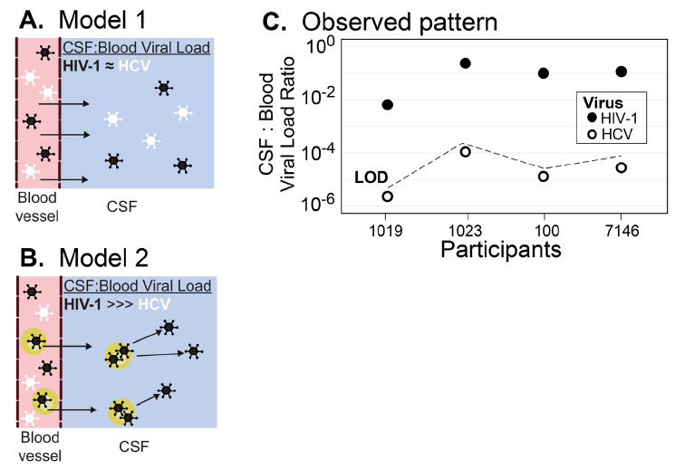
In this work, we carried out a cross-sectional study examining HIV-1 and HCV free virus concentrations in blood and cerebrospinal fluid (CSF) to determine whether HIV-1 enters the central nervous system (CNS) passively as virus particles or in the context of migrating infected cells. If virions migrate freely across the blood-cerebrospinal fluid barrier (BCSFB) or the blood-brain barrier (BBB) then HCV and HIV-1 would be detectable in the CSF at proportions similar to that in the blood. Alternatively, virus entry as an infected cell would favor selective entry of HIV-1.
We measured HIV-1 and HCV viral loads in the CSF and blood plasma of 4 co-infected participants who were not on antiviral regimens for either infection. We also generated HIV-1 sequences and performed phylogenetic analyses to determine whether HIV-1 populations in the CSF of these participants were being maintained by local replication.
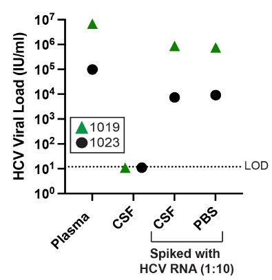
While CSF samples taken from all participants had detectable levels of HIV-1, HCV was not detectable in any of the CSF samples despite participants having HCV concentrations in their blood plasma, which exceeded that of HIV-1. Further, there was no evidence of compartmentalized HIV-1 replication in the CNS (Supplementary Figure 1). These results are consistent with a model where HIV-1 particles cross the BBB or the BCSFB within infected cells. In this scenario, we would expect HIV-1 to reach the CSF more readily because the blood contains a much greater number of HIV-infected cells than HCV-infected cells.
HCV entry into the CSF is restricted, indicating that virions do not freely migrate across these barriers and supporting the concept that HIV-1 is transported across the BCSFB and/or BBB by the migration of HIV-infected cells as part of an inflammatory response or normal surveillance.
在本研究中,我们开展了一项横断面研究,检测血液和脑脊液(CSF)中无HIV-1和HCV的病毒浓度,以确定HIV-1是以病毒颗粒的形式被动进入中枢神经系统(CNS),还是在感染细胞迁移的情况下进入。如果病毒粒子能自由穿过血脑脊液屏障(BCSFB)或血脑屏障(BBB),那么CSF中可检测到的HCV和HIV-1比例将与血液中的相似。另外,以感染细胞形式进入病毒则有利于HIV-1的选择性进入。
我们测量了4名同时感染两种病毒且未接受任何一种感染的抗病毒治疗方案的参与者的CSF和血浆中的HIV-1和HCV病毒载量。我们还生成了HIV-1序列并进行了系统发育分析,以确定这些参与者CSF中的HIV-1群体是否通过局部复制得以维持。
虽然所有参与者的CSF样本中都可检测到HIV-1水平,但尽管参与者血浆中的HCV浓度超过了HIV-1,所有CSF样本中均未检测到HCV。此外,没有证据表明CNS中存在HIV-1的分区复制(补充图1)。这些结果与HIV-1颗粒在感染细胞内穿过BBB或BCSFB的模型一致。在这种情况下,我们预计HIV-1更容易进入CSF,因为血液中HIV感染细胞的数量比HCV感染细胞多得多。
HCV进入CSF受到限制,这表明病毒粒子不会自由穿过这些屏障,并支持这样一种观点,即HIV-1作为炎症反应或正常监测的一部分,通过HIV感染细胞的迁移穿过BCSFB和/或BBB。