• 文献检索
  • 文档翻译
  • 深度研究
  • 学术资讯
  • Suppr Zotero 插件Zotero 插件
  • 邀请有礼
  • 套餐&价格
  • 历史记录
应用&插件
Suppr Zotero 插件Zotero 插件浏览器插件Mac 客户端Windows 客户端微信小程序
定价
高级版会员购买积分包购买API积分包
服务
文献检索文档翻译深度研究API 文档MCP 服务
关于我们
关于 Suppr公司介绍联系我们用户协议隐私条款
关注我们

Suppr 超能文献

核心技术专利:CN118964589B侵权必究
粤ICP备2023148730 号-1Suppr @ 2026

文献检索

告别复杂PubMed语法,用中文像聊天一样搜索,搜遍4000万医学文献。AI智能推荐,让科研检索更轻松。

立即免费搜索

文件翻译

保留排版,准确专业,支持PDF/Word/PPT等文件格式,支持 12+语言互译。

免费翻译文档

深度研究

AI帮你快速写综述,25分钟生成高质量综述,智能提取关键信息,辅助科研写作。

立即免费体验

青光眼的血管调节异常:视网膜血管收缩与高度性视野缺损中的正常神经血管耦合

Vascular dysregulation in glaucoma: retinal vasoconstriction and normal neurovascular coupling in altitudinal visual field defects.

作者信息

Zhou Wanshu, Sabel Bernhard A

机构信息

Institute of Medical Psychology, Medical Faculty, Otto-Von-Guericke University of Magdeburg, Leipziger Straße 44, 39120 Magdeburg, Germany.

出版信息

EPMA J. 2023 Feb 16;14(1):87-99. doi: 10.1007/s13167-023-00316-6. eCollection 2023 Mar.

DOI:10.1007/s13167-023-00316-6
PMID:36866155
原文链接:https://pmc.ncbi.nlm.nih.gov/articles/PMC9971397/
Abstract

PURPOSE

Vision loss in glaucoma is not only associated with elevated intraocular pressure and neurodegeneration, but vascular dysregulation (VD) is a major factor. To optimize therapy, an improved understanding of concepts of predictive, preventive, and personalized medicine (3PM) is needed which is based on a more detailed understanding of VD pathology. Specifically, to learn if the root cause of glaucomatous vision loss is of neuronal (degeneration) or vascular origin, we now studied neurovascular coupling (NVC) and vessel morphology and their relationship to vision loss in glaucoma.

METHODS

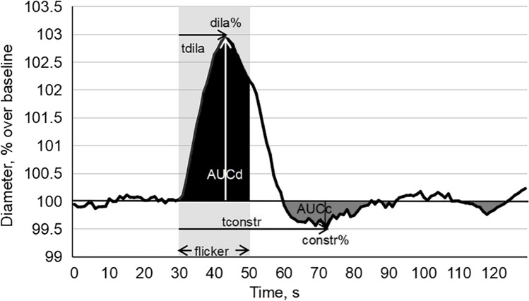
In patients with primary open angle glaucoma (POAG) ( = 30) and healthy controls ( = 22), NVC was studied using dynamic vessel analyzer to quantify retinal vessel diameter before, during, and after flicker light stimulation to evaluate the dilation response following neuronal activation. Vessel features and dilation were then related to branch level and visual field impairment.

RESULTS

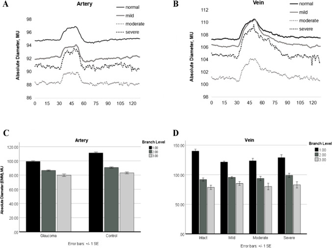
Retinal arterial and venous vessels had significantly smaller diameters in patients with POAG in comparison to controls. However, both arterial and venous dilation reached normal values during neuronal activation despite their smaller diameters. This was largely independent of visual field depth and varied among patients.

CONCLUSIONS

Because dilation/constriction is normal, VD in POAG can be explained by chronic vasoconstriction which limits energy supply to retinal (and brain) neurons with subsequent hypo-metabolism ("silent" neurons) or neuronal cell death. We propose that the root cause of POAG is primarily of vascular and not neuronal origin. This understanding can help to better personalize POAG therapy of not only targeting eye pressure but also vasoconstriction to prevent low vision, slowing its progression and supporting recovery and restoration.

TRIAL REGISTRATION

ClinicalTrials.gov, # NCT04037384 on July 3, 2019.

摘要

目的

青光眼导致的视力丧失不仅与眼压升高和神经退行性变有关,而且血管调节功能障碍(VD)也是一个主要因素。为了优化治疗,需要在更详细了解VD病理的基础上,更好地理解预测、预防和个性化医学(3PM)的概念。具体而言,为了弄清楚青光眼性视力丧失的根本原因是神经元(变性)还是血管源性,我们现在研究了神经血管耦合(NVC)和血管形态及其与青光眼视力丧失的关系。

方法

在原发性开角型青光眼(POAG)患者(n = 30)和健康对照者(n = 22)中,使用动态血管分析仪研究NVC,以量化闪烁光刺激前、期间和之后的视网膜血管直径,评估神经元激活后的扩张反应。然后将血管特征和扩张与分支水平和视野损害相关联。

结果

与对照组相比,POAG患者的视网膜动脉和静脉血管直径明显较小。然而,尽管它们的直径较小,但在神经元激活期间,动脉和静脉扩张均达到正常值。这在很大程度上与视野深度无关,且患者之间存在差异。

结论

由于扩张/收缩正常,POAG中的VD可以用慢性血管收缩来解释,慢性血管收缩限制了视网膜(和大脑)神经元的能量供应,随后出现代谢减退(“沉默”神经元)或神经元细胞死亡。我们提出,POAG的根本原因主要是血管源性而非神经元源性。这种认识有助于更好地对POAG治疗进行个性化,不仅针对眼压,还针对血管收缩,以预防低视力,减缓其进展并支持恢复和康复。

试验注册

ClinicalTrials.gov,# NCT04037384,2019年7月3日。

https://cdn.ncbi.nlm.nih.gov/pmc/blobs/121f/9971397/e748257df87a/13167_2023_316_Fig3_HTML.jpg
https://cdn.ncbi.nlm.nih.gov/pmc/blobs/121f/9971397/82b3328540d7/13167_2023_316_Fig1_HTML.jpg
https://cdn.ncbi.nlm.nih.gov/pmc/blobs/121f/9971397/d98553c8b1b1/13167_2023_316_Fig2_HTML.jpg
https://cdn.ncbi.nlm.nih.gov/pmc/blobs/121f/9971397/e748257df87a/13167_2023_316_Fig3_HTML.jpg
https://cdn.ncbi.nlm.nih.gov/pmc/blobs/121f/9971397/82b3328540d7/13167_2023_316_Fig1_HTML.jpg
https://cdn.ncbi.nlm.nih.gov/pmc/blobs/121f/9971397/d98553c8b1b1/13167_2023_316_Fig2_HTML.jpg
https://cdn.ncbi.nlm.nih.gov/pmc/blobs/121f/9971397/e748257df87a/13167_2023_316_Fig3_HTML.jpg

相似文献

1
Vascular dysregulation in glaucoma: retinal vasoconstriction and normal neurovascular coupling in altitudinal visual field defects.青光眼的血管调节异常:视网膜血管收缩与高度性视野缺损中的正常神经血管耦合
EPMA J. 2023 Feb 16;14(1):87-99. doi: 10.1007/s13167-023-00316-6. eCollection 2023 Mar.
2
Retinal neurovascular coupling in patients with glaucoma and ocular hypertension and its association with the level of glaucomatous damage.青光眼和高眼压症患者的视网膜神经血管耦合及其与青光眼损害程度的关系。
Graefes Arch Clin Exp Ophthalmol. 2013 Jun;251(6):1577-85. doi: 10.1007/s00417-013-2276-9. Epub 2013 Feb 19.
3
Does hemispheric vascular regulation differ significantly in glaucoma patients with altitudinal visual field asymmetry? A single-center, prospective study.高海拔视野不对称的青光眼患者的半球血管调节是否存在显著差异?一项单中心前瞻性研究。
Int Ophthalmol. 2021 Sep;41(9):3109-3119. doi: 10.1007/s10792-021-01876-0. Epub 2021 May 19.
4
Age-related impairment of neurovascular coupling responses: a dynamic vessel analysis (DVA)-based approach to measure decreased flicker light stimulus-induced retinal arteriolar dilation in healthy older adults.年龄相关性神经血管耦合反应损伤:一种基于动态血管分析(DVA)的方法,用于测量健康老年人闪烁光刺激诱导的视网膜小动脉扩张减少。
Geroscience. 2019 Jun;41(3):341-349. doi: 10.1007/s11357-019-00078-y. Epub 2019 Jun 17.
5
Loss of Caveolin-1 Impairs Light Flicker-Induced Neurovascular Coupling at the Optic Nerve Head.小窝蛋白-1缺失会损害视神经乳头处光闪烁诱导的神经血管耦合。
Front Neurosci. 2021 Nov 8;15:764898. doi: 10.3389/fnins.2021.764898. eCollection 2021.
6
[Retinal vessel density in primary open-angle glaucoma with a hemifield defect].[原发性开角型青光眼伴半视野缺损的视网膜血管密度]
Zhonghua Yan Ke Za Zhi. 2021 Mar 11;57(3):201-206. doi: 10.3760/cma.j.cn112142-20201102-00734.
7
Investigation of blood flow regulation and oxygen saturation of the retinal vessels in primary open-angle glaucoma.原发性开角型青光眼视网膜血管血流调节及血氧饱和度的研究
Graefes Arch Clin Exp Ophthalmol. 2014 Nov;252(11):1803-10. doi: 10.1007/s00417-014-2766-4. Epub 2014 Aug 12.
8
Dependence of diameters and oxygen saturation of retinal vessels on visual field damage and age in primary open-angle glaucoma.原发性开角型青光眼视网膜血管直径和血氧饱和度与视野损害及年龄的关系
Acta Ophthalmol. 2016 May;94(3):276-81. doi: 10.1111/aos.12727. Epub 2015 Apr 15.
9
Peripapillary and Macular Vessel Density in Patients with Primary Open-Angle Glaucoma and Unilateral Visual Field Loss.原发性开角型青光眼伴单侧视野缺损患者的视盘周围和黄斑血管密度。
Ophthalmology. 2018 Apr;125(4):578-587. doi: 10.1016/j.ophtha.2017.10.029. Epub 2017 Nov 22.
10
Vasoconstriction and Impairment of Neurovascular Coupling after Subarachnoid Hemorrhage: a Descriptive Analysis of Retinal Changes.蛛网膜下腔出血后的血管收缩和神经血管耦联障碍:视网膜变化的描述性分析。
Transl Stroke Res. 2018 Jun;9(3):284-293. doi: 10.1007/s12975-017-0585-8. Epub 2017 Nov 8.

引用本文的文献

1
Retinal Vessel Diameter Reductions Are Associated with Retinal Ganglion Cell Dysfunction, Thinning of the Ganglion Cell and Inner Plexiform Layers, and Decreased Visual Field Global Indices in Glaucoma Suspects.视网膜血管直径减小与青光眼可疑患者的视网膜神经节细胞功能障碍、神经节细胞层和内网状层变薄以及视野全局指标降低有关。
Diagnostics (Basel). 2025 Jul 3;15(13):1700. doi: 10.3390/diagnostics15131700.
2
Associations Between Cerebral Perfusion Pressure, Hemodynamic Parameters, and Cognitive Test Values in Normal-Tension Glaucoma Patients, Alzheimer's Disease Patients, and Healthy Controls.正常眼压性青光眼患者、阿尔茨海默病患者及健康对照者的脑灌注压、血流动力学参数与认知测试值之间的关联
Medicina (Kaunas). 2025 May 24;61(6):972. doi: 10.3390/medicina61060972.
3

本文引用的文献

1
Psychological interventions to reduce intraocular pressure (IOP) in glaucoma patients: a review.降低青光眼患者眼压的心理干预措施:一项综述
Graefes Arch Clin Exp Ophthalmol. 2023 May;261(5):1215-1227. doi: 10.1007/s00417-022-05912-2. Epub 2022 Nov 28.
2
cGMP Signaling in the Neurovascular Unit-Implications for Retinal Ganglion Cell Survival in Glaucoma.cGMP 信号在神经血管单元中的作用——对青光眼视网膜神经节细胞存活的影响。
Biomolecules. 2022 Nov 11;12(11):1671. doi: 10.3390/biom12111671.
3
Mitochondrial health quality control: measurements and interpretation in the framework of predictive, preventive, and personalized medicine.
Correlation of optic nerve hemoglobin levels with structural and functional parameters in glaucoma.青光眼患者视神经血红蛋白水平与结构和功能参数的相关性
Sci Rep. 2025 Jun 1;15(1):19190. doi: 10.1038/s41598-025-03549-9.
4
Comprehensive genome-wide analysis of retinal vessel caliber reveals microvascular-blood pressure pathways: advancing predictive, preventive, and personalized medicine.视网膜血管管径的全基因组综合分析揭示微血管-血压通路:推进预测性、预防性和个性化医学。
EPMA J. 2025 May 6;16(2):401-417. doi: 10.1007/s13167-025-00411-w. eCollection 2025 Jun.
5
Eye yoga for glaucoma: recovery of vascular dysregulation and visual field function-a randomized controlled trial.青光眼的眼瑜伽:血管调节异常和视野功能的恢复——一项随机对照试验
EPMA J. 2024 Dec 19;16(1):37-49. doi: 10.1007/s13167-024-00389-x. eCollection 2025 Mar.
6
Characterising the Time Course of the Dilatory Response of Healthy Retinal Arteries during Flicker-Light Provocation.描绘健康视网膜动脉在闪烁光刺激下扩张反应的时间进程。
J Vasc Res. 2025;62(1):1-9. doi: 10.1159/000541443. Epub 2024 Nov 19.
7
Genetic association of lipid traits and lipid-related drug targets with normal tension glaucoma: a Mendelian randomization study for predictive preventive and personalized medicine.脂质性状和脂质相关药物靶点与正常眼压性青光眼的遗传关联:一项用于预测性预防和个性化医学的孟德尔随机化研究
EPMA J. 2024 Jul 13;15(3):511-524. doi: 10.1007/s13167-024-00373-5. eCollection 2024 Sep.
8
Integrative neurovascular coupling and neurotransmitter analyses in anisometropic and visual deprivation amblyopia children.屈光参差性和视觉剥夺性弱视儿童的整合性神经血管耦合与神经递质分析
iScience. 2024 May 16;27(6):109988. doi: 10.1016/j.isci.2024.109988. eCollection 2024 Jun 21.
9
[Glaucoma Patient with Suspected Flammer Syndrome: Diagnostic Procedures and Therapeutic Implications].[疑似Flammer综合征的青光眼患者:诊断程序及治疗意义]
Klin Monbl Augenheilkd. 2024 Apr;241(4):355-360. doi: 10.1055/a-2275-2323. Epub 2024 Apr 23.
10
The Association between Vascular Abnormalities and Glaucoma-What Comes First?血管异常与青光眼之间的关联——哪个为先?
Int J Mol Sci. 2023 Aug 25;24(17):13211. doi: 10.3390/ijms241713211.
线粒体健康质量控制:预测、预防和个性化医学框架下的测量与解读
EPMA J. 2022 May 12;13(2):177-193. doi: 10.1007/s13167-022-00281-6. eCollection 2022 Jun.
4
Anxiety, depression, and sleep-related outcomes of glaucoma patients: systematic review and meta-analysis.青光眼患者的焦虑、抑郁和睡眠相关结果:系统评价和荟萃分析。
Can J Ophthalmol. 2023 Aug;58(4):346-355. doi: 10.1016/j.jcjo.2022.02.010. Epub 2022 Mar 16.
5
Pericyte dysfunction and loss of interpericyte tunneling nanotubes promote neurovascular deficits in glaucoma.周细胞功能障碍和周细胞间隧穿纳米管的缺失导致青光眼的神经血管缺损。
Proc Natl Acad Sci U S A. 2022 Feb 15;119(7). doi: 10.1073/pnas.2110329119.
6
Spacetime in the brain: rapid brain network reorganization in visual processing and recovery.大脑中的时空:视觉处理与恢复过程中快速的脑网络重组
Sci Rep. 2021 Sep 9;11(1):17940. doi: 10.1038/s41598-021-96971-8.
7
Endothelin-1 axes in the framework of predictive, preventive and personalised (3P) medicine.预测、预防和个性化(3P)医学框架下的内皮素-1轴
EPMA J. 2021 Aug 4;12(3):265-305. doi: 10.1007/s13167-021-00248-z. eCollection 2021 Sep.
8
The effect of daily life activities on intraocular pressure related variations in open-angle glaucoma.日常生活活动对开角型青光眼眼压相关变化的影响。
Sci Rep. 2021 Mar 23;11(1):6598. doi: 10.1038/s41598-021-85980-2.
9
Is Mental Stress the Primary Cause of Glaucoma?精神压力是青光眼的主要病因吗?
Klin Monbl Augenheilkd. 2021 Feb;238(2):132-145. doi: 10.1055/a-1303-8025. Epub 2021 Feb 12.
10
Neurovascular Unit: A critical role in ischemic stroke.神经血管单元:在缺血性脑卒中中的关键作用。
CNS Neurosci Ther. 2021 Jan;27(1):7-16. doi: 10.1111/cns.13561. Epub 2021 Jan 2.