Ophthalmology Department, Sohag Faculty of Medicine, Sohag University, Sohag, Egypt.
Ophthalmology Department, Faculty of Medicine, Al-Azhar University, Assiut, Egypt.
Indian J Ophthalmol. 2023 Mar;71(3):830-836. doi: 10.4103/ijo.IJO_1792_22.
To evaluate the use of Scheimpflug tomography in corneal densitometry (CD) in comparing the stages of keratoconic eyes.
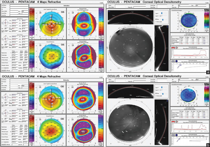
Keratoconic (KC) corneas (stages 1-3 classified according to the topographic parameters) were examined using the Scheimpflug tomographer (Pentacam, Oculus) using the CD software. CD was measured over three different depths (anterior stromal layer [120 μm], posterior stromal layer [60 μm], and middle stromal layer between these two layers), and concentric annular zones (0.0 to 2.0, 2.0 to 6.0, 6.0 to 10.0, and 10.0 to 12.0 mm diameter area).
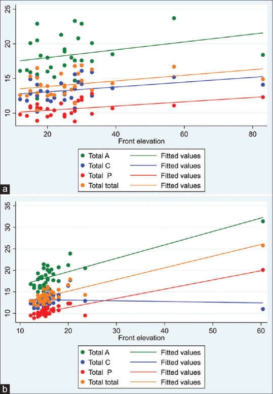
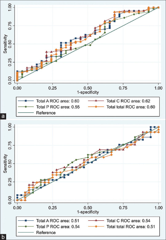
The study participants were divided into three groups: keratoconus (KC) stage 1 (KC1) with 64 participants, keratoconus stage 2 (KC2) with 29 participants, and keratoconus stage 3 (KC3) with 36 participants. Comparing CD of all three layers (anterior, central, and posterior) of the cornea over different circular annuli (0-2, 2-6, 6-10, and 10-12 mm) revealed a significant difference in the 6-10 mm annulus between all groups and in all layers (P = 0.3, 0.2, and 0.2, respectively). Area under curve (AUC) was done. It revealed that the central layer showed the highest specificity (93.8%) in comparing KC1 and KC2, whereas CD in the anterior layer between KC2 and KC3 had the highest specificity (86.2%).
CD showed increased values in the anterior corneal layer and in the annulus 6-10 mm more than other locations in all stages of KC.
评估 Scheimpflug 体层摄影术在角膜密度测量(CD)中用于比较圆锥角膜眼的阶段。
使用 Scheimpflug 断层扫描仪(Pentacam,Oculus)对角膜地形图参数分类为 1 至 3 期的圆锥角膜(KC)角膜进行检查,使用 CD 软件。在三个不同深度(前基质层[120μm]、后基质层[60μm]和两层之间的中间基质层)和同心环状区域(0.0 至 2.0、2.0 至 6.0、6.0 至 10.0 和 10.0 至 12.0mm 直径区域)测量 CD。
研究参与者分为三组:圆锥角膜 1 期(KC1)64 例,圆锥角膜 2 期(KC2)29 例,圆锥角膜 3 期(KC3)36 例。比较角膜所有三层(前、中、后)在不同圆形环(0-2、2-6、6-10 和 10-12mm)的 CD,发现所有组之间在 6-10mm 环和所有层之间的 CD 均有显著差异(分别为 P = 0.3、0.2 和 0.2)。进行了曲线下面积(AUC)分析。结果表明,中央层在比较 KC1 和 KC2 时具有最高的特异性(93.8%),而在 KC2 和 KC3 之间前层的 CD 具有最高的特异性(86.2%)。
在 KC 的所有阶段,CD 在角膜前层和 6-10mm 环处的测量值均高于其他部位。