Tuncer Zeynep, Dereli Can Gamze, Dönmez Keklikoğlu Hava, Eren Fatma Ayşen, Yülek Fatma, Deniz Orhan
Sakarya Adatıp Hospital, Neurology and Pain Clinic, Sakarya, Turkey.
Department of Ophthalmology, Bursa City Hospital, Bursa, Turkey.
Parkinsons Dis. 2023 Feb 23;2023:7739944. doi: 10.1155/2023/7739944. eCollection 2023.
In Parkinson's disease (PD), dopamine deficiency is present not only in the nigrostriatal pathway but also in the retinal and visual pathways. Optic coherence tomography (OCT) can be used as morphological evidence of visual influence from early nonmotor symptoms. The aim of this study was to investigate the relationship of OCT and visual evoked potentials (VEPs) of eyes with the severity of clinical findings and ocular findings in PD.
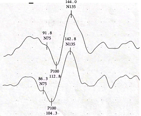
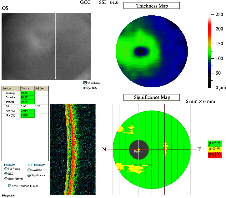
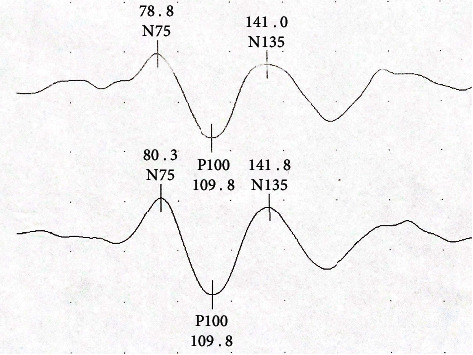
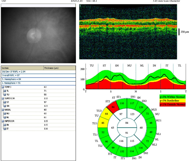
A group of 42 patients diagnosed with idiopathic PD and a control group of 29 people between the ages of 45-85 were included in our study. VEP was recorded in the patient and control groups. OCT measurement was made with the Optovue spectral-domain device. Foveal thickness and macular volume were measured in the foveal region and in the parafoveal and perifoveal regions in the temporal, superior, nasal, and inferior quadrants. RNFL (retinal nerve fiber layer) was measured in temporal, superior, nasal, and inferior quadrants. Ganglion cell complex (GCC) was evaluated in the superior and inferior quadrants. Using the UPDRS clinical scale, the relationship between measurements and the differences between the control group and the patient group were evaluated.
Among the OCT values in our study, foveal, parafoveal, perifoveal thickness, macular volume, RNFL, and GCC measurements were performed for the right and left eyes, and no difference was found between the patient group and the control group. There was no difference in VEP amplitude and latency values between the patient and control groups. The relationships between UPDRS and modified Hoehn Yahr staging and OCT and VEP measurements in the patient revealed no correlation.
Studies on whether OCT measurements can functionally be a marker or which segments are more valuable for disease progression in patients with PD are needed. Visual dysfunction in PD cannot be attributed only to retinal pathology; however, the retina may provide monitoring of the status of dopaminergic neurodegeneration and axonal loss in PD.
在帕金森病(PD)中,多巴胺缺乏不仅存在于黑质纹状体通路,还存在于视网膜和视觉通路。光学相干断层扫描(OCT)可作为早期非运动症状对视觉影响的形态学证据。本研究旨在探讨PD患者眼部的OCT和视觉诱发电位(VEP)与临床及眼部检查结果严重程度之间的关系。
本研究纳入了一组42例诊断为特发性PD的患者以及一个由29名年龄在45 - 85岁之间的人组成的对照组。对患者组和对照组进行了VEP记录。使用Optovue光谱域设备进行OCT测量。在黄斑区以及颞侧、上方、鼻侧和下方象限的旁黄斑区和黄斑周围区域测量黄斑厚度和黄斑体积。在颞侧、上方、鼻侧和下方象限测量视网膜神经纤维层(RNFL)。在上方和下方象限评估神经节细胞复合体(GCC)。使用统一帕金森病评定量表(UPDRS)临床量表,评估测量值与对照组和患者组之间差异之间的关系。
在本研究的OCT值中,对左右眼进行了黄斑、旁黄斑、黄斑周围厚度以及黄斑体积、RNFL和GCC测量,患者组和对照组之间未发现差异。患者组和对照组之间的VEP振幅和潜伏期值也没有差异。患者的UPDRS与改良Hoehn - Yahr分期以及OCT和VEP测量之间的关系未显示出相关性。
需要开展研究以确定OCT测量在功能上是否可作为一个标志物,或者对于PD患者疾病进展而言哪些节段更具价值。PD中的视觉功能障碍不能仅归因于视网膜病变;然而,视网膜可能有助于监测PD中多巴胺能神经变性和轴突丢失的状态。