• 文献检索
  • 文档翻译
  • 深度研究
  • 学术资讯
  • Suppr Zotero 插件Zotero 插件
  • 邀请有礼
  • 套餐&价格
  • 历史记录
应用&插件
Suppr Zotero 插件Zotero 插件浏览器插件Mac 客户端Windows 客户端微信小程序
定价
高级版会员购买积分包购买API积分包
服务
文献检索文档翻译深度研究API 文档MCP 服务
关于我们
关于 Suppr公司介绍联系我们用户协议隐私条款
关注我们

Suppr 超能文献

核心技术专利:CN118964589B侵权必究
粤ICP备2023148730 号-1Suppr @ 2026

文献检索

告别复杂PubMed语法,用中文像聊天一样搜索,搜遍4000万医学文献。AI智能推荐,让科研检索更轻松。

立即免费搜索

文件翻译

保留排版,准确专业,支持PDF/Word/PPT等文件格式,支持 12+语言互译。

免费翻译文档

深度研究

AI帮你快速写综述,25分钟生成高质量综述,智能提取关键信息,辅助科研写作。

立即免费体验

使用Q-FIX MINI缝线锚钉进行内镜下腓骨上支持带重建术

Endoscopic Superior Peroneal Retinaculum Reconstruction Using Q-FIX MINI Suture Anchor.

作者信息

Lui Tun Hing, Li Charles Churk Hang

机构信息

Department of Orthopaedics and Traumatology, Hong Kong SAR, China.

North District Hospital, Hong Kong SAR, China.

出版信息

Arthrosc Tech. 2023 Jan 18;12(2):e233-e240. doi: 10.1016/j.eats.2022.11.001. eCollection 2023 Feb.

DOI:10.1016/j.eats.2022.11.001
PMID:36879874
原文链接:https://pmc.ncbi.nlm.nih.gov/articles/PMC9984787/
Abstract

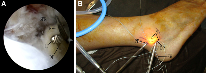
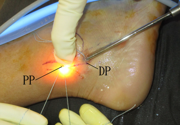
Post-traumatic peroneal tendon subluxation or dislocation is most commonly caused by injury to the superior peroneal retinaculum. Classic open surgeries usually require extensive soft-tissue dissection and have potential risks of peritendinous fibrous adhesions, sural nerve injury, limited range of movement, recurrent or persistent peroneal tendon instability, and tendon irritation. The purpose of this Technical Note is to describe the details of endoscopic superior peroneal retinaculum reconstruction using Q-FIX MINI suture anchor. This endoscopic approach has the advantages of minimally invasive surgery, including better cosmesis, less soft-tissue dissection, less postoperative pain, less peritendinous fibrosis, and less subjective tightness at peroneal tendons. Insertion of the Q-FIX MINI suture anchor can be performed inside a drill guide, and trapping of the surrounding soft tissue can be avoided.

摘要

创伤后腓骨肌腱半脱位或脱位最常见的原因是腓骨上支持带损伤。传统的开放手术通常需要广泛的软组织分离,并且存在腱周纤维粘连、腓肠神经损伤、活动范围受限、腓骨肌腱反复或持续不稳定以及肌腱刺激等潜在风险。本技术说明的目的是描述使用Q-FIX MINI缝合锚进行内镜下腓骨上支持带重建的细节。这种内镜方法具有微创手术的优点,包括更好的美容效果、更少的软组织分离、术后疼痛减轻、腱周纤维化减少以及腓骨肌腱处主观紧绷感减轻。Q-FIX MINI缝合锚可以在钻孔导向器内插入,并且可以避免周围软组织的卡压。

https://cdn.ncbi.nlm.nih.gov/pmc/blobs/06c8/9984787/3356b6935f8c/gr10.jpg
https://cdn.ncbi.nlm.nih.gov/pmc/blobs/06c8/9984787/beda4701ba15/gr1.jpg
https://cdn.ncbi.nlm.nih.gov/pmc/blobs/06c8/9984787/503dbf35bdae/gr2.jpg
https://cdn.ncbi.nlm.nih.gov/pmc/blobs/06c8/9984787/3b0f81968fbf/gr3.jpg
https://cdn.ncbi.nlm.nih.gov/pmc/blobs/06c8/9984787/9d90b1dbbb8a/gr4.jpg
https://cdn.ncbi.nlm.nih.gov/pmc/blobs/06c8/9984787/0ff9a3f11605/gr5.jpg
https://cdn.ncbi.nlm.nih.gov/pmc/blobs/06c8/9984787/8c737d95940b/gr6.jpg
https://cdn.ncbi.nlm.nih.gov/pmc/blobs/06c8/9984787/336a34df00aa/gr7.jpg
https://cdn.ncbi.nlm.nih.gov/pmc/blobs/06c8/9984787/3ef7dcb7ffdb/gr8.jpg
https://cdn.ncbi.nlm.nih.gov/pmc/blobs/06c8/9984787/c23cea5e939c/gr9.jpg
https://cdn.ncbi.nlm.nih.gov/pmc/blobs/06c8/9984787/3356b6935f8c/gr10.jpg

相似文献

1
Endoscopic Superior Peroneal Retinaculum Reconstruction Using Q-FIX MINI Suture Anchor.使用Q-FIX MINI缝线锚钉进行内镜下腓骨上支持带重建术
Arthrosc Tech. 2023 Jan 18;12(2):e233-e240. doi: 10.1016/j.eats.2022.11.001. eCollection 2023 Feb.
2
Endoscopic Superior Peroneal Retinaculum Reconstruction.内镜下腓骨上支持带重建术。
Arthrosc Tech. 2017 Dec 18;7(1):e45-e51. doi: 10.1016/j.eats.2017.08.050. eCollection 2018 Jan.
3
Knotless Tendoscopic Peroneal Retinaculum Repair Technique for Recurrent Peroneal Tendon Dislocation.用于复发性腓骨肌腱脱位的无结关节镜下腓骨支持带修复技术
Arthrosc Tech. 2022 Jul 14;11(8):e1395-e1401. doi: 10.1016/j.eats.2022.03.028. eCollection 2022 Aug.
4
Eckert and Davis Grade 3 Superior Peroneal Retinaculum Injury: Treated by Endoscopic Peroneal Retinaculum Reconstruction and Complicated by Malposition of the Suture Anchors.埃克特和戴维斯3级腓骨上支持带损伤:经内镜下腓骨支持带重建治疗,并发缝合锚钉位置不当。
J Orthop Case Rep. 2015 Oct-Dec;5(4):73-6. doi: 10.13107/jocr.2250-0685.352.
5
Treatment of Recurrent Peroneal Tendon Dislocation by Peroneal Retinaculum Reattachment Without Fibular Groove Deepening.腓骨肌腱沟不加深的腓骨肌腱支持带修复治疗复发性腓骨肌腱脱位
J Foot Ankle Surg. 2021 Sep-Oct;60(5):994-997. doi: 10.1053/j.jfas.2020.04.027. Epub 2021 Jun 18.
6
Peroneal Stabilization Via Tightening of the Peroneal Tendon Sheath.通过拉紧腓肠肌腱鞘实现腓骨稳定。
Foot Ankle Spec. 2022 Dec;15(6):566-572. doi: 10.1177/19386400211068240. Epub 2022 Jan 12.
7
Reconstruction of the superior peroneal retinaculum using an autologous gracilis tendon graft for chronic dislocation of the peroneal tendons accompanied by lateral instability of the ankle: technical note.使用自体股薄肌腱移植重建腓骨上支持带治疗伴有踝关节外侧不稳定的腓骨肌腱慢性脱位:技术说明
Knee Surg Sports Traumatol Arthrosc. 2007 Apr;15(4):461-4. doi: 10.1007/s00167-006-0258-8. Epub 2006 Dec 23.
8
Snapping ankles: peroneal tendon subluxation and dislocation.踝关节扭伤:腓骨肌腱脱位和脱位。
Br J Hosp Med (Lond). 2023 Oct 2;84(10):1-7. doi: 10.12968/hmed.2022.0257. Epub 2023 Sep 29.
9
Tendoscopic Double-Row Suture Bridge Peroneal Retinaculum Repair for Recurrent Dislocation of Peroneal Tendons in the Ankle.关节镜下双排缝线桥修复术治疗踝关节腓骨肌腱复发性脱位
Arthrosc Tech. 2016 May 2;5(3):e441-6. doi: 10.1016/j.eats.2016.01.028. eCollection 2016 Jun.
10
Superior peroneal retinaculum reattachment for an atraumatic peroneus brevis tendon subluxation: a case report.高位腓骨支持带修复术治疗非创伤性腓骨短肌腱半脱位:病例报告。
J Med Case Rep. 2022 Jun 16;16(1):239. doi: 10.1186/s13256-022-03455-y.

引用本文的文献

1
Recurrent peroneal tendon dislocation-the current concept of management.复发性腓骨肌腱脱位——当前的治疗理念
Ann Jt. 2024 Aug 30;9:40. doi: 10.21037/aoj-24-10. eCollection 2024.
2
Comparison of Clinical Efficacy between Arthroscopic and Open Surgery for Ogden Type 1-2 Peroneal Tendon Dislocation.关节镜与开放手术治疗 Ogden 1-2 型腓骨肌腱脱位的临床疗效比较。
Orthop Surg. 2024 May;16(5):1079-1088. doi: 10.1111/os.14035. Epub 2024 Mar 21.

本文引用的文献

1
Treatment of Recurrent Peroneal Tendon Dislocation by Peroneal Retinaculum Reattachment Without Fibular Groove Deepening.腓骨肌腱沟不加深的腓骨肌腱支持带修复治疗复发性腓骨肌腱脱位
J Foot Ankle Surg. 2021 Sep-Oct;60(5):994-997. doi: 10.1053/j.jfas.2020.04.027. Epub 2021 Jun 18.
2
Concomitant Lateral Ankle and Peroneal Tendon Instability Treated With Simultaneous Arthroscopic Broström and Tendoscopic Superior Peroneal Retinaculum Repair: A Case Report.关节镜下 Broström 术与经皮内窥镜下上支持带修复同期治疗合并的外踝和腓骨肌腱不稳定:病例报告。
J Foot Ankle Surg. 2021 Jul-Aug;60(4):850-855. doi: 10.1053/j.jfas.2021.01.005. Epub 2021 Feb 9.
3
Reattachment of the superior peroneal retinaculum versus the bone block procedure for the treatment of recurrent peroneal tendon dislocation: two safe and effective techniques.
修复上支持带与骨块固定术治疗复发性外踝肌腱脱位:两种安全有效的技术。
Knee Surg Sports Traumatol Arthrosc. 2019 Sep;27(9):2877-2883. doi: 10.1007/s00167-019-05479-2. Epub 2019 Mar 22.
4
Endoscopic Superior Peroneal Retinaculum Reconstruction.内镜下腓骨上支持带重建术。
Arthrosc Tech. 2017 Dec 18;7(1):e45-e51. doi: 10.1016/j.eats.2017.08.050. eCollection 2018 Jan.
5
Eckert and Davis Grade 3 Superior Peroneal Retinaculum Injury: Treated by Endoscopic Peroneal Retinaculum Reconstruction and Complicated by Malposition of the Suture Anchors.埃克特和戴维斯3级腓骨上支持带损伤:经内镜下腓骨支持带重建治疗,并发缝合锚钉位置不当。
J Orthop Case Rep. 2015 Oct-Dec;5(4):73-6. doi: 10.13107/jocr.2250-0685.352.
6
Functional outcomes after peroneal tendoscopy in the treatment of peroneal tendon disorders.腓骨肌腱镜检查治疗腓骨肌腱疾病后的功能结果。
Knee Surg Sports Traumatol Arthrosc. 2016 Apr;24(4):1148-54. doi: 10.1007/s00167-016-4012-6. Epub 2016 Feb 4.
7
Peroneus Longus Tendoscopy at the Sole: A Cadaveric Study.足底腓骨长肌腱镜检查:一项尸体研究。
Arthroscopy. 2015 Jul;31(7):1338-42. doi: 10.1016/j.arthro.2015.01.024. Epub 2015 Mar 19.
8
Endoscopic management of recalcitrant retrofibular pain without peroneal tendon subluxation or dislocation.内镜治疗顽固性腓骨后疼痛而无腓骨肌腱脱位或半脱位。
Arch Orthop Trauma Surg. 2012 Mar;132(3):357-61. doi: 10.1007/s00402-011-1392-4. Epub 2011 Sep 10.
9
A preliminary report on intra-sheath peroneal tendon subluxation: a prospective review of 7 patients with ultrasound verification.鞘内腓骨肌腱半脱位的初步报告:7例超声证实患者的前瞻性研究
J Foot Ankle Surg. 2009 May-Jun;48(3):323-9. doi: 10.1053/j.jfas.2009.02.003.
10
Retromalleolar groove impaction for the treatment of unstable peroneal tendons.后踝沟嵌压术治疗不稳定的腓骨肌腱
Am J Sports Med. 2009 Jan;37(1):191-4. doi: 10.1177/0363546508324310. Epub 2008 Oct 16.