Department of Gynaecology and Obstetrics, The First Affiliate Hospital of Jinan University, Jinan University, No.613 Huangpu Road West, Guangzhou, 510632, China.
International Joint Laboratory for Embryonic Development & Prenatal Medicine, Division of Histology and Embryology, Medical College, Jinan University, Guangzhou, 510632, China.
BMC Med. 2023 Mar 9;21(1):90. doi: 10.1186/s12916-023-02807-9.
Pre-eclampsia (PE) is one of the leading causes of maternal and fetal morbidity/mortality during pregnancy, and alpha-2-macroglobulin (A2M) is associated with inflammatory signaling; however, the pathophysiological mechanism by which A2M is involved in PE development is not yet understood.
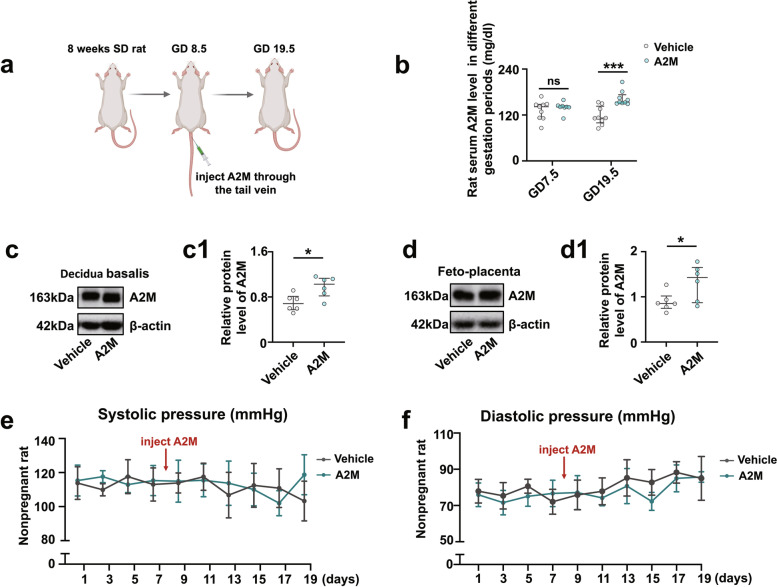
Human placenta samples, serum, and corresponding clinical data of the participants were collected to study the pathophysiologic mechanism underlying PE. Pregnant Sprague-Dawley rats were intravenously injected with an adenovirus vector carrying A2M via the tail vein on gestational day (GD) 8.5. Human umbilical artery smooth muscle cells (HUASMCs), human umbilical vein endothelial cells (HUVECs), and HTR-8/SVneo cells were transfected with A2M-expressing adenovirus vectors.
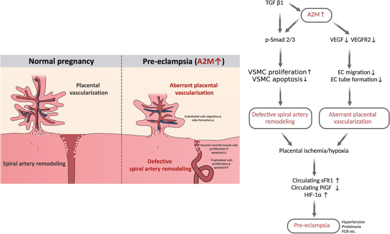
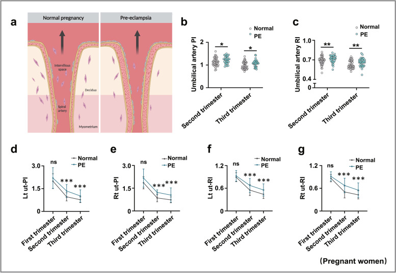
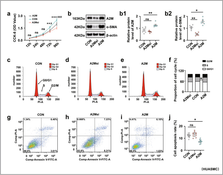
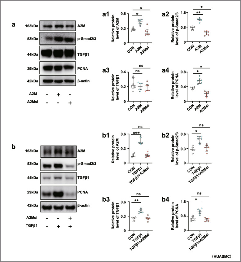
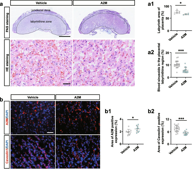
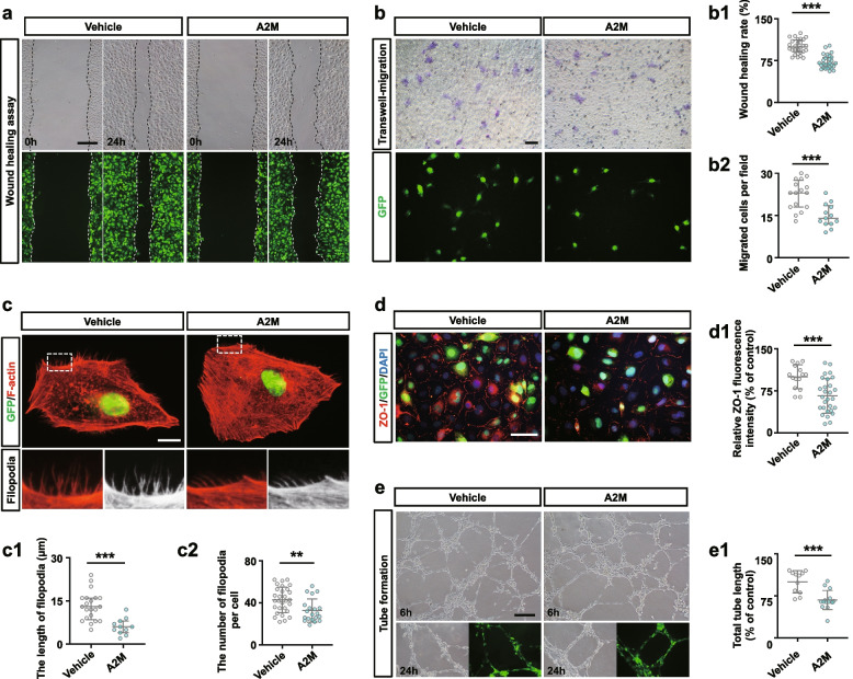
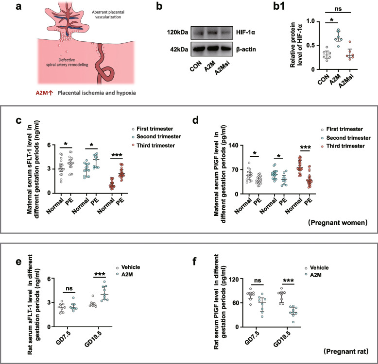
In this study, we demonstrated that A2M levels were significantly increased in PE patient serum, uterine spiral arteries, and feto-placental vasculature. The A2M-overexpression rat model closely mimicked the characteristics of PE (i.e., hypertension in mid-to-late gestation, histological and ultrastructural signs of renal damage, proteinuria, and fetal growth restriction). Compared to the normal group, A2M overexpression significantly enhanced uterine artery vascular resistance and impaired uterine spiral artery remodeling in both pregnant women with early-onset PE and in pregnant rats. We found that A2M overexpression was positively associated with HUASMC proliferation and negatively correlated with cell apoptosis. In addition, the results demonstrated that transforming growth factor beta 1 (TGFβ1) signaling regulated the effects of A2M on vascular muscle cell proliferation described above. Meanwhile, A2M overexpression regressed rat placental vascularization and reduced the expression of angiogenesis-related genes. In addition, A2M overexpression reduced HUVEC migration, filopodia number/length, and tube formation. Furthermore, HIF-1α expression was positively related to A2M, and the secretion of sFLT-1 and PIGF of placental origin was closely related to PE during pregnancy or A2M overexpression in rats.
Our data showed that gestational A2M overexpression can be considered a contributing factor leading to PE, causing detective uterine spiral artery remodeling and aberrant placental vascularization.
子痫前期(PE)是妊娠期间母婴发病率和死亡率的主要原因之一,α-2-巨球蛋白(A2M)与炎症信号有关;然而,A2M 参与 PE 发展的病理生理机制尚不清楚。
收集参与者的人胎盘样本、血清和相应的临床数据,以研究 PE 的病理生理机制。妊娠 Sprague-Dawley 大鼠于妊娠第 8.5 天经尾静脉静脉注射携带 A2M 的腺病毒载体。转染 A2M 表达腺病毒载体的人脐动脉平滑肌细胞(HUASMCs)、人脐静脉内皮细胞(HUVECs)和 HTR-8/SVneo 细胞。
在这项研究中,我们证明了 A2M 水平在 PE 患者血清、子宫螺旋动脉和胎儿胎盘血管中显著增加。A2M 过表达大鼠模型密切模拟了 PE 的特征(即中晚期妊娠高血压、肾损伤的组织学和超微结构特征、蛋白尿和胎儿生长受限)。与正常组相比,A2M 过表达显着增加了子宫动脉血管阻力,并损害了早发型 PE 孕妇和妊娠大鼠的子宫螺旋动脉重塑。我们发现 A2M 过表达与 HUASMC 增殖呈正相关,与细胞凋亡呈负相关。此外,结果表明转化生长因子β 1(TGFβ1)信号调节了 A2M 对上述血管平滑肌细胞增殖的影响。同时,A2M 过表达逆转了大鼠胎盘血管化并降低了血管生成相关基因的表达。此外,A2M 过表达减少了 HUVEC 迁移、丝状伪足数量/长度和管形成。此外,HIF-1α 表达与 A2M 呈正相关,胎盘来源的 sFLT-1 和 PIGF 的分泌与妊娠期间的 PE 或大鼠中的 A2M 过表达密切相关。
我们的数据表明,妊娠期间 A2M 过表达可被认为是导致 PE 的一个促成因素,导致子宫螺旋动脉重塑不良和胎盘血管化异常。