Otsuka Yasuo, Komeda Yoriaki, Takeda Masayuki, Takahama Takayuki, Kono Masashi, Takenaka Mamoru, Hagiwara Satoru, Nishida Naoshi, Kashida Hiroshi, Kudo Masatoshi
Department of Gastroenterology and Hepatology, Kindai University Faculty of Medicine, Osaka-Sayama, Japan.
Department of Cancer Genomics and Medical Oncology, Nara Medical University, 840 Shijo-Cho, Kashihara, Nara, Japan.
Case Rep Med. 2023 Feb 28;2023:2092157. doi: 10.1155/2023/2092157. eCollection 2023.
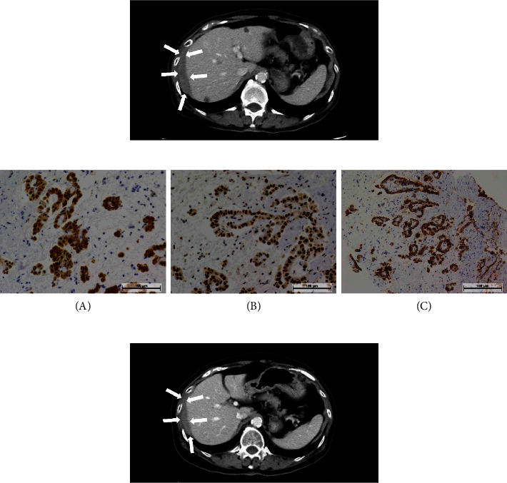
A 76-year-old woman presented with lower abdominal pain and nausea and was referred to the gastroenterology department in our institution. Previous contrast-enhanced computed tomography (CE-CT) for follow-up after breast cancer surgery had indicated a soft tissue mass below the right diaphragm, which was considered a benign change. CE-CT performed at the first visit to our department revealed further thickening of the soft tissue mass with extension to the liver surface. In addition, ascites and nodules were observed in the abdominal cavity. Histopathological examination of a biopsy specimen revealed peritoneal invasion of atypical epithelioid cells with trabecular and glandular patterns. The tumor cells were positive for AE1/AE2, calretinin, WT-1, D2-40, HEG1, EMA, BAP1, and MTAP and negative for carcinoembryonic antigen, MOC-31, Ber-Ep4, ER, PgR, TTF-1, claudin 4, and desmin. A diagnosis of epithelioid mesothelioma was made. The patient received chemotherapy with cisplatin (75 mg/m) and pemetrexed (500 mg/m). After six courses of combined chemotherapy, pemetrexed was administered as a single agent. At the time of writing this report, she was undergoing over the 30th course of chemotherapy without any significant side effects. Diffuse malignant peritoneal mesothelioma is a rare, fatal, and progressive disease. Our patient achieved long-term survival of more than 5 years with maintenance therapy using single-agent pemetrexed.
一名76岁女性因下腹部疼痛和恶心前来就诊,被转诊至我院胃肠病科。此前乳腺癌手术后进行的对比增强计算机断层扫描(CE-CT)显示右膈下有一软组织肿块,当时认为是良性改变。在我院初诊时进行的CE-CT显示软组织肿块进一步增厚,并延伸至肝表面。此外,腹腔内观察到腹水和结节。活检标本的组织病理学检查显示非典型上皮样细胞呈小梁状和腺管状模式侵犯腹膜。肿瘤细胞AE1/AE2、钙视网膜蛋白、WT-1、D2-40、HEG1、EMA、BAP1和MTAP呈阳性,癌胚抗原、MOC-31、Ber-Ep4、ER、PgR、TTF-1、claudin 4和结蛋白呈阴性。诊断为上皮样间皮瘤。患者接受了顺铂(75mg/m)和培美曲塞(500mg/m)化疗。联合化疗六个疗程后,改为培美曲塞单药治疗。在撰写本报告时,她正在接受第30多个疗程的化疗,未出现任何明显副作用。弥漫性恶性腹膜间皮瘤是一种罕见、致命且进行性的疾病。我们的患者通过培美曲塞单药维持治疗实现了超过5年的长期生存。