Nakamura Masatoshi, Ohnishi Kayoko, Uchida Fumihiko, Saito Takashi, Kitagawa Yuri, Matsuoka Ryota, Yanagawa Toru, Sakurai Hideyuki
Department of Radiation Oncology, Faculty of Medicine, University of Tsukuba, 1-1-1 Tennodai, Tsukuba, Ibaraki 305-8577 Japan.
Department of Radiology, School of Medicine, International University of Health and Welfare, 4-3 Kozunomori, Narita, Chiba 286-8686 Japan.
Int Cancer Conf J. 2023 Feb 1;12(2):160-165. doi: 10.1007/s13691-023-00597-8. eCollection 2023 Apr.
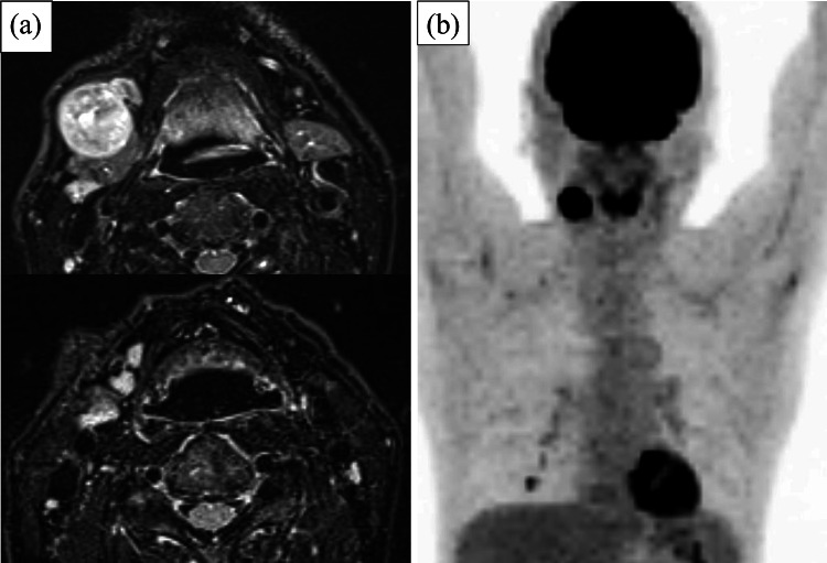
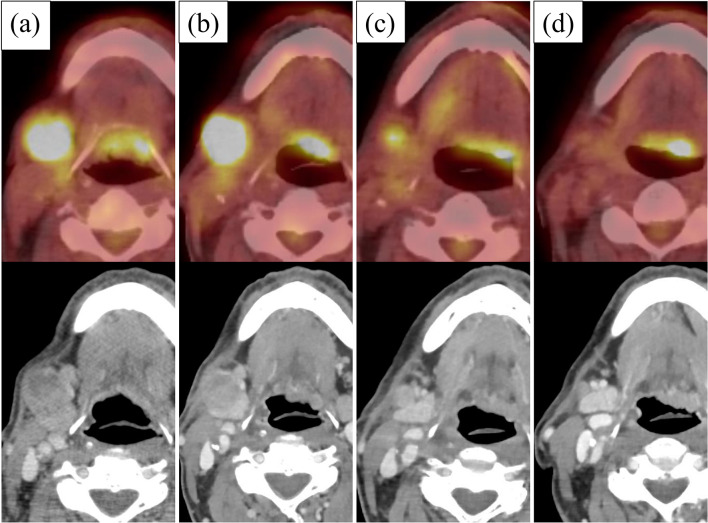
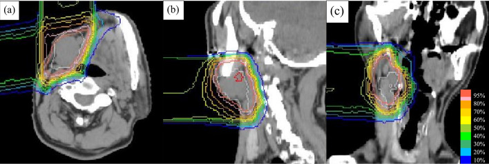
An 80-year-old man with an approximately 3-cm mass in the right submandibular region presented to our institution. Magnetic resonance imaging revealed enlarged lymph nodes (LNs) in the right neck, and fluorine-18-2-deoxy-D-glucose (FDG) positron emission tomography (PET)/computed tomography (CT) indicated positive FDG accumulation in the right neck LNs only. Excisional biopsy was performed for suspected malignant lymphoma, and the biopsy revealed melanoma. Close examination of the skin, nasal cavity, oral pharyngeal and laryngeal cavities, and gastrointestinal tract were performed. No primary tumor was detected by these examinations, and the patient was diagnosed with cervical LN metastasis from melanoma of unknown primary of clinical stage T0N3bM0 stage IIIC. The patient refused cervical neck dissection because of his age and comorbidity of Alzheimer's disease and instead opted for proton beam therapy (PBT) at a total dose of 69 Gy (relative biological effectiveness) in 23 fractions. He did not receive any systemic therapy. The enlarged LNs shrunk slowly, and FDG PET/CT at 1 year after PBT showed that the right submandibular LN had shrunk from 27 to 7 mm in length, and there was no significant FDG accumulation. At 6 years and 4 months after PBT, the patient is alive without any recurrence.
一名80岁男性因右下颌下区有一约3厘米肿物前来我院就诊。磁共振成像显示右侧颈部淋巴结肿大,氟-18-2-脱氧-D-葡萄糖(FDG)正电子发射断层扫描(PET)/计算机断层扫描(CT)仅显示右侧颈部淋巴结有FDG摄取阳性。对疑似恶性淋巴瘤进行了切除活检,结果显示为黑色素瘤。对皮肤、鼻腔、口咽和喉腔以及胃肠道进行了仔细检查。这些检查未发现原发肿瘤,该患者被诊断为临床分期为T0N3bM0 IIIC期的原发灶不明的黑色素瘤颈部淋巴结转移。由于患者年龄及合并阿尔茨海默病,拒绝行颈部淋巴结清扫术,而是选择了质子束治疗(PBT),总剂量为69 Gy(相对生物效应),分23次给予。他未接受任何全身治疗。肿大的淋巴结缓慢缩小,PBT后1年的FDG PET/CT显示右下颌下淋巴结长度从27毫米缩小至7毫米,且无明显FDG摄取。PBT后6年4个月,患者存活,无任何复发。