Department of Radiology, Weill Cornell Medical College, New York, NY, USA.
Department of Radiology, Shandong Provincial Hospital Affiliated to Shandong First Medical University, Jinan, Shandong, China.
Korean J Radiol. 2023 Apr;24(4):324-337. doi: 10.3348/kjr.2022.0652. Epub 2023 Mar 7.
The objective of this study was to analyze the different brain oxygen metabolism statuses in preeclampsia using magnetic resonance imaging and investigate the factors that affect cerebral oxygen metabolism in preeclampsia.
Forty-nine women with preeclampsia (mean age 32.4 years; range, 18-44 years), 22 pregnant healthy controls (PHCs) (mean age 30.7 years; range, 23-40 years), and 40 non-pregnant healthy controls (NPHCs) (mean age 32.5 years; range, 20-42 years) were included in this study. Brain oxygen extraction fraction (OEF) values were computed using quantitative susceptibility mapping (QSM) plus quantitative blood oxygen level-dependent magnitude-based OEF mapping (QSM + quantitative blood oxygen level-dependent imaging or QQ) obtained with a 1.5-T scanner. Voxel-based morphometry (VBM) was used to investigate the differences in OEF values in the brain regions among the groups.
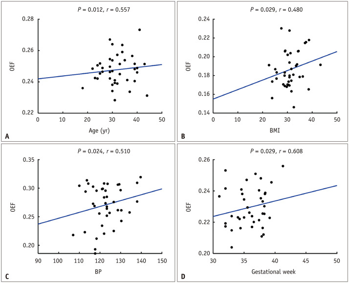
Among the three groups, the average OEF values were significantly different in multiple brain areas, including the parahippocampus, multiple gyri of the frontal lobe, calcarine, cuneus, and precuneus (all -values were less than 0.05, after correcting for multiple comparisons). The average OEF values of the preeclampsia group were higher than those of the PHC and NPHC groups. The bilateral superior frontal gyrus/bilateral medial superior frontal gyrus had the largest size of the aforementioned brain regions, and the OEF values in this area were 24.2 ± 4.6, 21.3 ± 2.4, and 20.6 ± 2.8 in the preeclampsia, PHC, and NPHC groups, respectively. In addition, the OEF values showed no significant differences between NPHC and PHC. Correlation analysis revealed that the OEF values of some brain regions (mainly involving the frontal, occipital, and temporal gyrus) were positively correlated with age, gestational week, body mass index, and mean blood pressure in the preeclampsia group ( = 0.361-0.812).
Using whole-brain VBM analysis, we found that patients with preeclampsia had higher OEF values than controls.
本研究旨在通过磁共振成像分析子痫前期患者不同的脑氧代谢状态,并探讨影响子痫前期脑氧代谢的因素。
本研究纳入了 49 例子痫前期患者(平均年龄 32.4 岁;范围 18-44 岁)、22 例正常妊娠健康对照组(PHC)(平均年龄 30.7 岁;范围 23-40 岁)和 40 例非妊娠健康对照组(NPHC)(平均年龄 32.5 岁;范围 20-42 岁)。使用定量磁化率映射(QSM)加上定量血氧水平依赖幅度 OEF 映射(QSM+定量血氧水平依赖成像或 QQ)在 1.5-T 扫描仪上计算脑氧摄取分数(OEF)值。体素形态学分析(VBM)用于研究三组之间脑区 OEF 值的差异。
三组之间,多个脑区的平均 OEF 值存在显著差异,包括海马旁回、额叶多个脑回、距状回、楔前叶和楔叶(所有 - 值均小于 0.05,校正多重比较后)。子痫前期组的平均 OEF 值高于 PHC 组和 NPHC 组。双侧额上回/双侧额上回内侧的脑区最大,OEF 值分别为子痫前期、PHC 和 NPHC 组的 24.2±4.6、21.3±2.4 和 20.6±2.8。此外,NPHC 组与 PHC 组之间的 OEF 值无显著差异。相关性分析显示,子痫前期组部分脑区(主要涉及额、顶、颞叶)的 OEF 值与年龄、孕周、体质量指数和平均血压呈正相关( = 0.361-0.812)。
使用全脑 VBM 分析,我们发现子痫前期患者的 OEF 值高于对照组。