Memudu Adejoke Elizabeth, Dongo Gambo A
Department of Anatomy, Faculty of Basic Medical Sciences, College of Medical Sciences, Edo State University Uzairue, Edo State Nigeria.
Department of Anatomy, Faculty of Basic Medical Sciences, Bingham University Karu Nasarawa State Nigeria.
Toxicol Rep. 2023 Feb 28;10:320-326. doi: 10.1016/j.toxrep.2023.02.010. eCollection 2023.
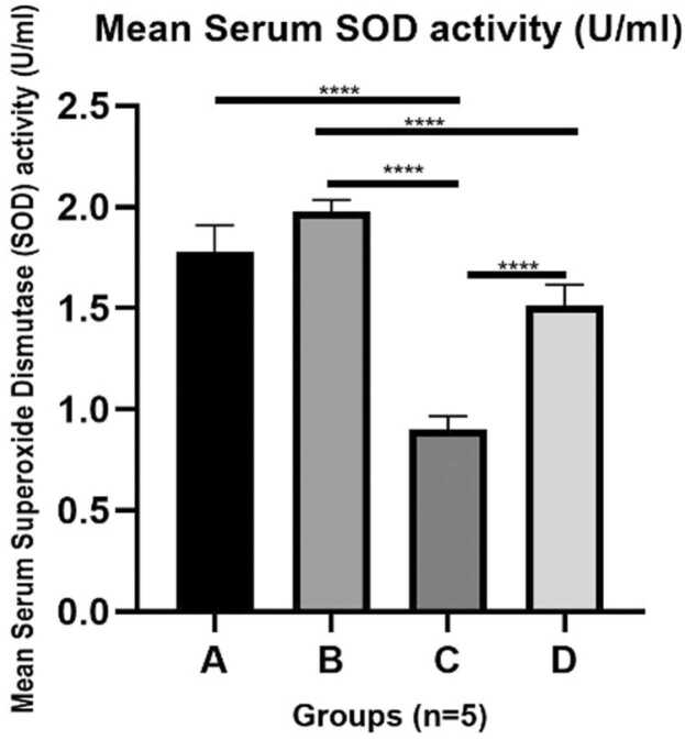
Anabolic Androgenic steroids (AAS) are abused and reports have been made on their deleterious effects on various organs. It is imperative to report the mechanism of inducing oxidative tissue damage even in the presence of an intracellular antioxidant system by the interaction between lipid peroxidation and the antioxidant system in the kidney. Twenty (20) adult male Wistar rats used were grouped into: A- Control, BOlive oil vehicle, C- 120 mg/kg of AAS orally for three weeks, and D- 7 days withdrawal group following 120 mg/kg/ 21days of AAS intake. Serum was assayed for lipid peroxidation marker Malondialdehyde (MDA) and antioxidant enzyme -superoxide Dismutase (SOD). Sectioned of kidneys were stained to see the renal tissue, mucin granules, and basement membrane. AAS-induced oxidative tissue damage, in the presence of an endogenous antioxidant, is characterized by increased lipid peroxidation and decreased SOD level which resulted in the loss of renal tissue cells membrane integrity which is a characteristic of the pathophysiology of nephron toxicity induced by a toxic compound. However, this was progressively reversed by a period of discontinuation of AAS drug exposure.
合成代谢雄激素类固醇(AAS)被滥用,且已有关于其对各种器官有害影响的报道。即使在存在细胞内抗氧化系统的情况下,通过肾脏中脂质过氧化与抗氧化系统之间的相互作用来报告诱导氧化组织损伤的机制也很有必要。所使用的20只成年雄性Wistar大鼠被分为:A组——对照组,B组——橄榄油载体组,C组——口服120mg/kg的AAS,持续三周,以及D组——在摄入120mg/kg/21天的AAS后停药7天的组。检测血清中的脂质过氧化标志物丙二醛(MDA)和抗氧化酶超氧化物歧化酶(SOD)。对肾脏切片进行染色以观察肾组织、粘蛋白颗粒和基底膜。在存在内源性抗氧化剂的情况下,AAS诱导的氧化组织损伤的特征是脂质过氧化增加和SOD水平降低,这导致肾组织细胞膜完整性丧失,这是有毒化合物诱导的肾单位毒性病理生理学的一个特征。然而,通过一段时间停止AAS药物暴露,这种情况逐渐得到逆转。