Massachusetts General Hospital Department of Neurology, Harvard Medical School, Boston, Massachusetts 02129, United States.
Advanced Proteomics Facility, Department of Biochemistry, University of Oxford, Oxford, Oxfordshire OX1 3QU, United Kingdom.
J Am Soc Mass Spectrom. 2023 Apr 5;34(4):649-667. doi: 10.1021/jasms.2c00341. Epub 2023 Mar 13.
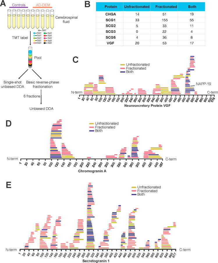
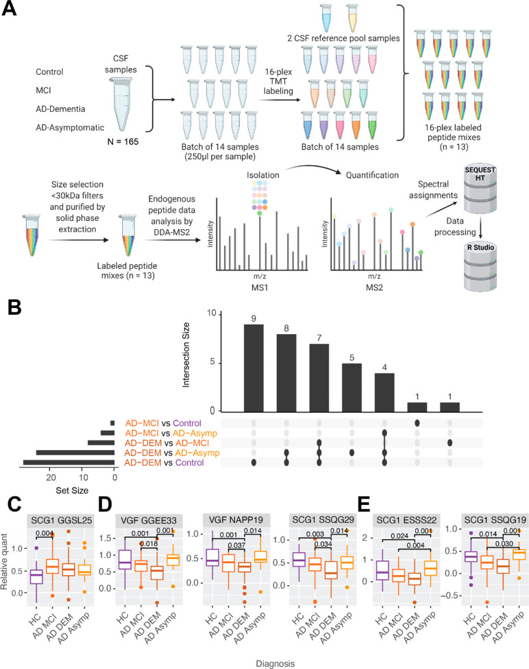
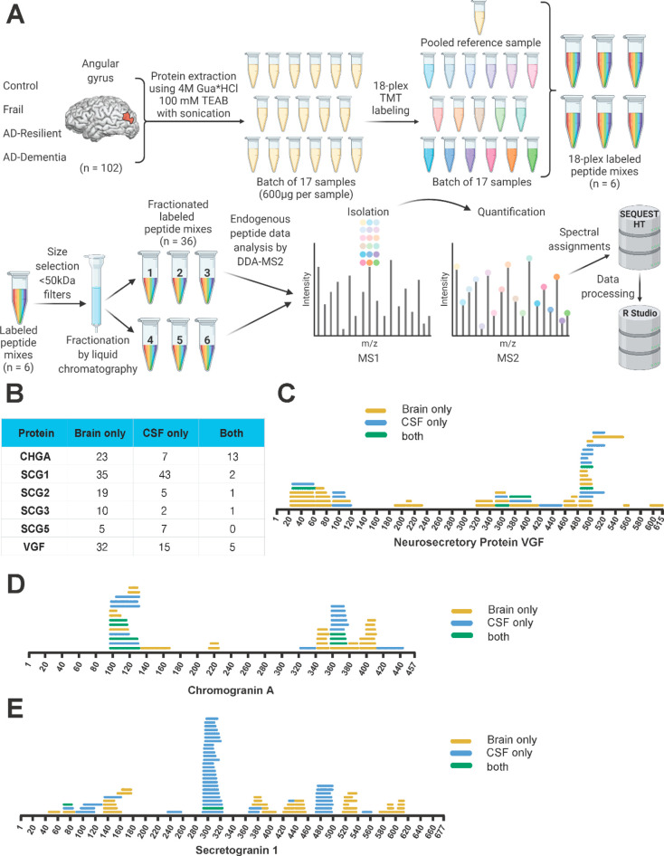
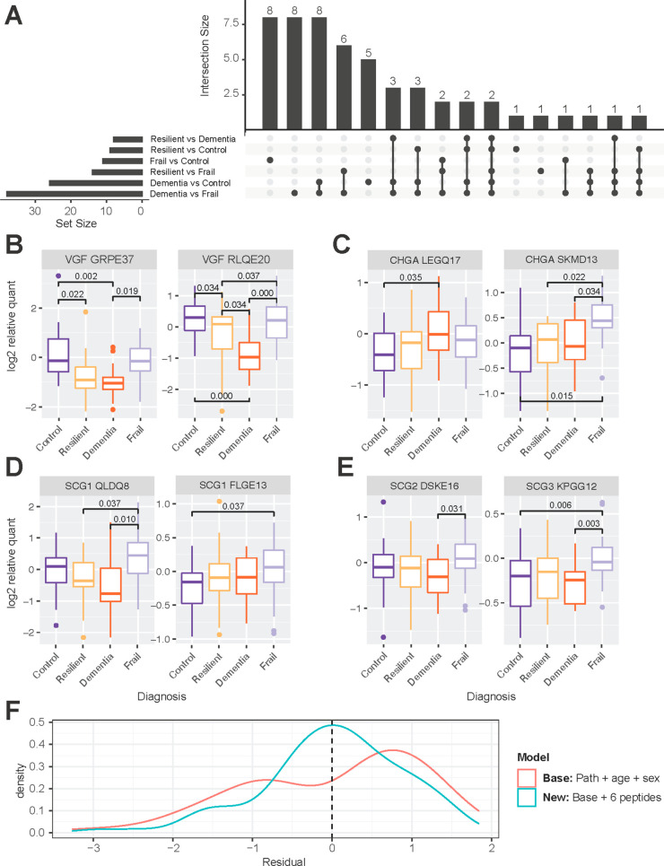
The granin neuropeptide family is composed of acidic secretory signaling molecules that act throughout the nervous system to help modulate synaptic signaling and neural activity. Granin neuropeptides have been shown to be dysregulated in different forms of dementia, including Alzheimer's disease (AD). Recent studies have suggested that the granin neuropeptides and their protease-cleaved bioactive peptides (proteoforms) may act as both powerful drivers of gene expression and as a biomarker of synaptic health in AD. The complexity of granin proteoforms in human cerebrospinal fluid (CSF) and brain tissue has not been directly addressed. We developed a reliable nontryptic mass spectrometry assay to comprehensively map and quantify endogenous neuropeptide proteoforms in the brain and CSF of individuals diagnosed with mild cognitive impairment and dementia due to AD compared to healthy controls, individuals with preserved cognition despite AD pathology ("Resilient"), and those with impaired cognition but no AD or other discernible pathology ("Frail"). We drew associations between neuropeptide proteoforms, cognitive status, and AD pathology values. Decreased levels of VGF proteoforms were observed in CSF and brain tissue from individuals with AD compared to controls, while select proteoforms from chromogranin A showed the opposite effect. To address mechanisms of neuropeptide proteoform regulation, we showed that the proteases Calpain-1 and Cathepsin S can cleave chromogranin A, secretogranin-1, and VGF into proteoforms found in both the brain and CSF. We were unable to demonstrate differences in protease abundance in protein extracts from matched brains, suggesting that regulation may occur at the level of transcription.
颗粒蛋白神经肽家族由酸性分泌信号分子组成,这些分子在整个神经系统中发挥作用,有助于调节突触信号和神经活动。颗粒蛋白神经肽已被证明在不同形式的痴呆症中失调,包括阿尔茨海默病 (AD)。最近的研究表明,颗粒蛋白神经肽及其蛋白酶切割的生物活性肽 (蛋白水解物) 可能既是基因表达的强大驱动因素,也是 AD 中突触健康的生物标志物。颗粒蛋白水解物在人脑脊液 (CSF) 和脑组织中的复杂性尚未得到直接解决。我们开发了一种可靠的非胰蛋白酶质谱测定法,用于全面绘制和定量个体诊断为轻度认知障碍和 AD 引起的痴呆与健康对照者、AD 病理但认知保留的个体(“Resilient”)以及认知受损但无 AD 或其他可识别病理的个体(“Frail”)的大脑和 CSF 中的内源性神经肽蛋白水解物。我们将神经肽蛋白水解物与认知状态和 AD 病理学值之间的关联。与对照组相比,AD 患者的 CSF 和脑组织中观察到 VGF 蛋白水解物水平降低,而来自嗜铬粒蛋白 A 的某些蛋白水解物则显示出相反的效果。为了解决神经肽蛋白水解物调节的机制,我们表明钙蛋白酶-1 和组织蛋白酶 S 可以将嗜铬粒蛋白 A、分泌颗粒蛋白-1 和 VGF 切割成在大脑和 CSF 中都发现的蛋白水解物。我们无法证明来自匹配大脑的蛋白质提取物中蛋白酶丰度的差异,这表明调节可能发生在转录水平。