• 文献检索
  • 文档翻译
  • 深度研究
  • 学术资讯
  • Suppr Zotero 插件Zotero 插件
  • 邀请有礼
  • 套餐&价格
  • 历史记录
应用&插件
Suppr Zotero 插件Zotero 插件浏览器插件Mac 客户端Windows 客户端微信小程序
定价
高级版会员购买积分包购买API积分包
服务
文献检索文档翻译深度研究API 文档MCP 服务
关于我们
关于 Suppr公司介绍联系我们用户协议隐私条款
关注我们

Suppr 超能文献

核心技术专利:CN118964589B侵权必究
粤ICP备2023148730 号-1Suppr @ 2026

文献检索

告别复杂PubMed语法,用中文像聊天一样搜索,搜遍4000万医学文献。AI智能推荐,让科研检索更轻松。

立即免费搜索

文件翻译

保留排版,准确专业,支持PDF/Word/PPT等文件格式,支持 12+语言互译。

免费翻译文档

深度研究

AI帮你快速写综述,25分钟生成高质量综述,智能提取关键信息,辅助科研写作。

立即免费体验

成年大鼠肾被膜下培养的生殖结节中阴茎骨的发育。

Development of the os penis in genital tubercles cultured beneath the renal capsule of adult rats.

作者信息

Murakami R

机构信息

Zoological Institute, Faculty of Science, University of Tokyo, Japan.

出版信息

J Anat. 1986 Dec;149:11-20.

PMID:3693100
原文链接:https://pmc.ncbi.nlm.nih.gov/articles/PMC1261630/
Abstract

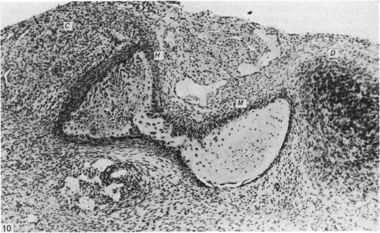
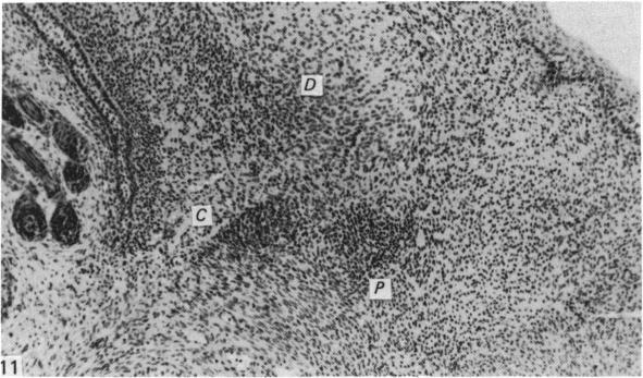
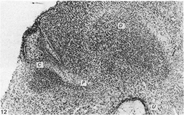
Genital tubercles of male and female rats were cultured beneath the renal capsule of castrated and intact adult male rats treated with androgens, oestrogen, or anti-androgen, and the development of the os penis in the transplants was studied. When the genital tubercles were cultured in normal male hosts, a membrane bone and a hyaline cartilage of the proximal segment of the os penis were formed 8-11 days after transplantation, and a fibrocartilage of the distal segment of the os penis at 11-14 days. In genital tubercles cultured in castrated males, the rudiments of both the proximal and distal segments remained as undifferentiated mesenchymal cell masses. However, similarly cultured genital tubercles were found to develop cartilages and bone when the hosts were treated with high doses of androgens. The potency of androgen-dependent chondrogenesis and osteogenesis was equivalent in the male and female genital tubercles. Chondrogenesis and osteogenesis of the os penis were caused by androgens, while the rudiments of the os penis were formed independently of androgens. The overt differentiation of the corpus cavernosum penis was also caused by androgens.

摘要

将雄性和雌性大鼠的生殖结节移植到经雄激素、雌激素或抗雄激素处理的去势成年雄性大鼠和完整成年雄性大鼠的肾被膜下,研究移植组织中阴茎骨的发育情况。当生殖结节在正常雄性宿主中培养时,移植后8 - 11天形成阴茎骨近端的膜性骨和透明软骨,11 - 14天形成阴茎骨远端的纤维软骨。在去势雄性大鼠中培养的生殖结节,近端和远端的原基均保持为未分化的间充质细胞团。然而,当宿主用高剂量雄激素处理时,同样培养的生殖结节会发育出软骨和骨。雄激素依赖性软骨生成和成骨能力在雄性和雌性生殖结节中相当。阴茎骨的软骨生成和成骨是由雄激素引起的,而阴茎骨的原基是独立于雄激素形成的。阴茎海绵体的明显分化也是由雄激素引起的。

https://cdn.ncbi.nlm.nih.gov/pmc/blobs/df0e/1261630/09d966f191cb/janat00183-0023-c.jpg
https://cdn.ncbi.nlm.nih.gov/pmc/blobs/df0e/1261630/c28455e70c8e/janat00183-0020-a.jpg
https://cdn.ncbi.nlm.nih.gov/pmc/blobs/df0e/1261630/6d01574c3b0e/janat00183-0020-b.jpg
https://cdn.ncbi.nlm.nih.gov/pmc/blobs/df0e/1261630/231f42f04b4d/janat00183-0020-c.jpg
https://cdn.ncbi.nlm.nih.gov/pmc/blobs/df0e/1261630/b3543d645566/janat00183-0020-d.jpg
https://cdn.ncbi.nlm.nih.gov/pmc/blobs/df0e/1261630/55180d1e8705/janat00183-0021-a.jpg
https://cdn.ncbi.nlm.nih.gov/pmc/blobs/df0e/1261630/689810f4dfd3/janat00183-0021-b.jpg
https://cdn.ncbi.nlm.nih.gov/pmc/blobs/df0e/1261630/227e2b2b7f10/janat00183-0021-c.jpg
https://cdn.ncbi.nlm.nih.gov/pmc/blobs/df0e/1261630/deb485ec17d1/janat00183-0021-d.jpg
https://cdn.ncbi.nlm.nih.gov/pmc/blobs/df0e/1261630/75c4b725261f/janat00183-0022-a.jpg
https://cdn.ncbi.nlm.nih.gov/pmc/blobs/df0e/1261630/273af57a8862/janat00183-0023-a.jpg
https://cdn.ncbi.nlm.nih.gov/pmc/blobs/df0e/1261630/94dc42286a4f/janat00183-0023-b.jpg
https://cdn.ncbi.nlm.nih.gov/pmc/blobs/df0e/1261630/09d966f191cb/janat00183-0023-c.jpg

相似文献

1
Development of the os penis in genital tubercles cultured beneath the renal capsule of adult rats.成年大鼠肾被膜下培养的生殖结节中阴茎骨的发育。
J Anat. 1986 Dec;149:11-20.
2
Proximal-distal sequence of development of the skeletal tissues in the penis of rat and the inductive effect of epithelium.大鼠阴茎骨骼组织发育的近远侧序列及上皮的诱导作用
J Embryol Exp Morphol. 1986 Mar;92:133-43.
3
Autoradiographic studies of the localisation of androgen-binding cells in the genital tubercles of fetal rats.胎鼠生殖结节中雄激素结合细胞定位的放射自显影研究。
J Anat. 1987 Apr;151:209-19.
4
Androgen-dependent and independent process of bone formation in the distal segment of Os penis in the rat.大鼠阴茎骨远端雄激素依赖和非依赖的骨形成过程。
Eur J Morphol. 1995 Nov;33(4):393-400.
5
[Organotypic culture of the genital tubercle of the fetal rat: induction of the os penis by testosterone].[胎鼠生殖结节的器官型培养:睾酮诱导阴茎骨形成]
C R Seances Soc Biol Fil. 1984;178(5):576-9.
6
A histological study of the development of the penis of wild-type and androgen-insensitive mice.野生型和雄激素不敏感小鼠阴茎发育的组织学研究。
J Anat. 1987 Aug;153:223-31.
7
The effect of neonatal treatment of male mice with antiandrogens and of females with androgens on the development of the os penis and os clitoridis.用抗雄激素对雄性小鼠进行新生期处理以及用雄激素对雌性小鼠进行新生期处理,对阴茎骨和阴蒂骨发育的影响。
J Anat. 1976 Apr;121(Pt 2):363-70.
8
Ontogeny of androgen receptor and disruption of its mRNA expression by exogenous estrogens during morphogenesis of the genital tubercle.雄激素受体的个体发生以及在生殖结节形态发生过程中外源雌激素对其mRNA表达的破坏。
J Urol. 2006 Oct;176(4 Pt 2):1883-8. doi: 10.1016/S0022-5347(06)00613-6.
9
Ultrastructure of the developing fibrocartilage of the os penis of rat.大鼠阴茎骨发育中纤维软骨的超微结构
J Morphol. 2000 Feb;243(2):187-91. doi: 10.1002/(SICI)1097-4687(200002)243:2<187::AID-JMOR7>3.0.CO;2-4.
10
Histogenesis of the Os Penis and Os Clitoridis in Rats: (chondrogenesis/bone formation/testosterone/genital tubercle).大鼠阴茎骨和阴蒂骨的组织发生:(软骨形成/骨形成/睾酮/生殖结节)
Dev Growth Differ. 1984;26(5):419-426. doi: 10.1111/j.1440-169X.1984.00419.x.

引用本文的文献

1
Genetic disruption of the baculum compromises the ability of male mice to copulate.阴茎骨的基因破坏会损害雄性小鼠的交配能力。
PLoS Genet. 2025 Jul 16;21(7):e1011787. doi: 10.1371/journal.pgen.1011787. eCollection 2025 Jul.
2
Comparative Morphology of the Penis and Clitoris in Four Species of Moles (Talpidae).四种鼹鼠(鼹科)阴茎与阴蒂的比较形态学
J Exp Zool B Mol Dev Evol. 2017 May;328(3):275-294. doi: 10.1002/jez.b.22732. Epub 2017 Mar 2.
3
Sonic hedgehog delivery from self-assembled nanofiber hydrogels reduces the fibrotic response in models of erectile dysfunction.

本文引用的文献

1
Testosterone, dihydrotestosterone and androstanediols in plasma, testes and prostates of rats during development.
Acta Endocrinol (Copenh). 1981 Jan;96(1):127-35. doi: 10.1530/acta.0.0960127.
2
Prostatic induction: interaction of epithelium and mesenchyme from normal wild-type mice and androgen-insensitive mice with testicular feminization.前列腺诱导:正常野生型小鼠和患有睾丸女性化的雄激素不敏感小鼠的上皮与间充质的相互作用。
J Endocrinol. 1980 Jun;85(3):423-8. doi: 10.1677/joe.0.0850423.
3
Os penis of the rat. IV. The proximal growth cartilage.大鼠阴茎骨。IV. 近端生长软骨。
从自组装纳米纤维水凝胶中递送音猬因子可降低勃起功能障碍模型中的纤维化反应。
Acta Biomater. 2016 Mar 1;32:89-99. doi: 10.1016/j.actbio.2016.01.014. Epub 2016 Jan 14.
4
New insights on the morphology of adult mouse penis.成年鼠阴茎形态的新认识。
Biol Reprod. 2011 Dec;85(6):1216-21. doi: 10.1095/biolreprod.111.091504. Epub 2011 Sep 14.
5
Estrogen-induced developmental disorders of the rat penis involve both estrogen receptor (ESR)- and androgen receptor (AR)-mediated pathways.雌激素诱导的大鼠阴茎发育障碍涉及雌激素受体 (ESR) 和雄激素受体 (AR) 介导的途径。
Biol Reprod. 2009 Sep;81(3):507-16. doi: 10.1095/biolreprod.108.071951. Epub 2009 May 6.
6
Autoradiographic studies of the localisation of androgen-binding cells in the genital tubercles of fetal rats.胎鼠生殖结节中雄激素结合细胞定位的放射自显影研究。
J Anat. 1987 Apr;151:209-19.
7
A histological study of the development of the penis of wild-type and androgen-insensitive mice.野生型和雄激素不敏感小鼠阴茎发育的组织学研究。
J Anat. 1987 Aug;153:223-31.
8
Fibrocartilage.纤维软骨
J Anat. 1990 Aug;171:1-15.
Acta Anat (Basel). 1983;117(2):136-44. doi: 10.1159/000145779.
4
Testosterone, dihydrotestosterone and estradiol-17 beta levels in maternal and fetal plasma and in fetal testes in the rat.大鼠母体和胎儿血浆以及胎儿睾丸中睾酮、双氢睾酮和雌二醇-17β水平
J Steroid Biochem. 1984 Aug;21(2):193-8. doi: 10.1016/0022-4731(84)90383-2.
5
[Organotypic culture of the genital tubercle of the fetal rat: induction of the os penis by testosterone].[胎鼠生殖结节的器官型培养:睾酮诱导阴茎骨形成]
C R Seances Soc Biol Fil. 1984;178(5):576-9.
6
Involution and hormone-induced persistence of the M. sphincter (levator) ani in female rats.雌性大鼠肛门括约肌(提肌)的退化及激素诱导的持续性变化
J Anat. 1970 Jan;106(Pt 1):93-110.
7
The hormonal induction of an os clitoridis in the neonatal and adult rat.新生和成年大鼠阴蒂骨的激素诱导作用。
J Anat. 1972 Jul;112(Pt 2):223-31.
8
Testosterone in the fetal rat testis.
Biol Reprod. 1973 Jun;8(5):560-5. doi: 10.1093/biolreprod/8.5.560.
9
Proximal-distal sequence of development of the skeletal tissues in the penis of rat and the inductive effect of epithelium.大鼠阴茎骨骼组织发育的近远侧序列及上皮的诱导作用
J Embryol Exp Morphol. 1986 Mar;92:133-43.
10
Tissue interaction in androgen response of embryonic mammary rudiment of mouse: identification of target tissue for testosterone.小鼠胚胎乳腺原基雄激素反应中的组织相互作用:睾酮靶组织的鉴定。
Proc Natl Acad Sci U S A. 1976 Nov;73(11):4041-4. doi: 10.1073/pnas.73.11.4041.