Shahidehpour Ryan K, Nelson Abraham S, Sanders Lydia G, Embry Chloe R, Nelson Peter T, Bachstetter Adam D
Spinal Cord and Brain Injury Research Center, University of Kentucky, 741 S. Limestone St., Lexington, KY, 40536, USA.
Department of Neuroscience, University of Kentucky, Lexington, KY, 40536, USA.
Acta Neuropathol Commun. 2023 Mar 18;11(1):45. doi: 10.1186/s40478-023-01541-w.
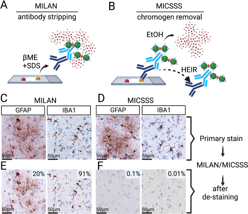
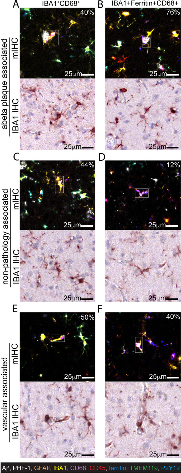
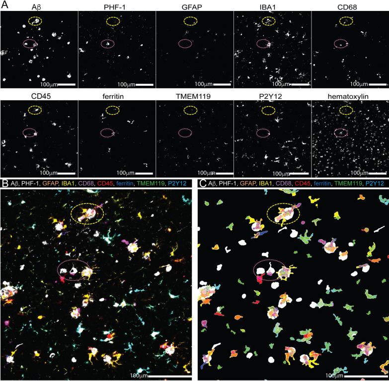
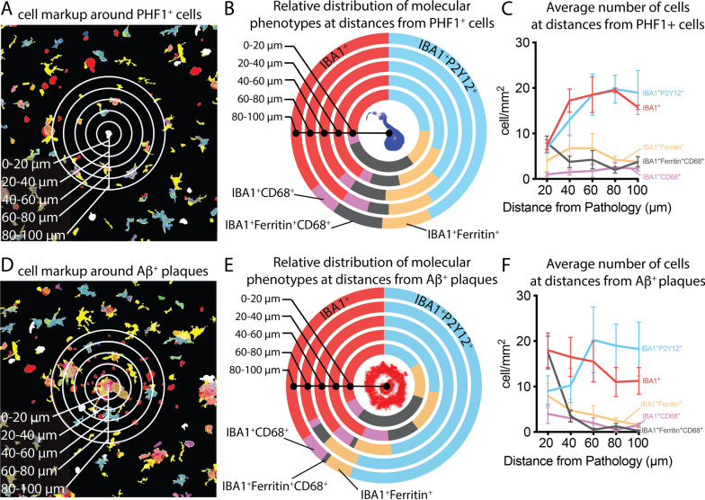
New histological techniques are needed to examine protein distribution in human tissues, which can reveal cell shape and disease pathology connections. Spatial proteomics has changed the study of tumor microenvironments by identifying spatial relationships of immunomodulatory cells and proteins and contributing to the discovery of new cancer immunotherapy biomarkers. However, the fast-expanding toolkit of spatial proteomic approaches has yet to be systematically applied to investigate pathological alterations in the aging human brain in health and disease states. Moreover, post-mortem human brain tissue presents distinct technical problems due to fixation procedures and autofluorescence, which limit fluorescence methodologies. This study sought to develop a multiplex immunohistochemistry approach (visualizing the immunostain with brightfield microscopy). Quantitative multiplex Immunohistochemistry with Visual colorimetric staining to Enhance Regional protein localization (QUIVER) was developed to address these technical challenges. Using QUIVER, a ten-channel pseudo-fluorescent image was generated using chromogen removal and digital microscopy to identify unique molecular microglia phenotypes. Next, the study asked if the tissue environment, specifically the amyloid plaques and neurofibrillary tangles characteristic of Alzheimer's disease, has any bearing on microglia's cellular and molecular phenotypes. QUIVER allowed the visualization of five molecular microglia/macrophage phenotypes using digital pathology tools. The recognizable reactive and homeostatic microglia/macrophage phenotypes demonstrated spatial polarization towards and away from amyloid plaques, respectively. Yet, microglia morphology appearance did not always correspond to molecular phenotype. This research not only sheds light on the biology of microglia but also offers QUIVER, a new tool for examining pathological alterations in the brains of the elderly.
需要新的组织学技术来检查人体组织中的蛋白质分布,这可以揭示细胞形状与疾病病理学之间的联系。空间蛋白质组学通过识别免疫调节细胞和蛋白质的空间关系,改变了肿瘤微环境的研究,并有助于发现新的癌症免疫治疗生物标志物。然而,快速扩展的空间蛋白质组学方法工具包尚未系统地应用于研究健康和疾病状态下衰老人类大脑的病理变化。此外,由于固定程序和自发荧光,死后人类脑组织存在明显的技术问题,这限制了荧光方法的应用。本研究旨在开发一种多重免疫组织化学方法(用明场显微镜观察免疫染色)。开发了定量多重免疫组织化学与视觉比色染色以增强区域蛋白质定位(QUIVER)来应对这些技术挑战。使用QUIVER,通过发色团去除和数字显微镜生成了一个十通道伪荧光图像,以识别独特的分子小胶质细胞表型。接下来,该研究询问组织环境,特别是阿尔茨海默病特有的淀粉样斑块和神经原纤维缠结,是否对小胶质细胞的细胞和分子表型有任何影响。QUIVER允许使用数字病理学工具可视化五种分子小胶质细胞/巨噬细胞表型。可识别的反应性和稳态小胶质细胞/巨噬细胞表型分别显示出朝向和远离淀粉样斑块的空间极化。然而,小胶质细胞的形态外观并不总是与分子表型相对应。这项研究不仅揭示了小胶质细胞的生物学特性,还提供了QUIVER,这是一种用于检查老年人脑部病理变化的新工具。