Mei Kai, Geagan Michael, Shapira Nadav, Liu Leening P, Pasyar Pouyan, Gang Grace J, Stayman J Webster, Noël Peter B
Department of Radiology, Perelman School of Medicine, University of Pennsylvania, Philadelphia, PA, USA.
Department of Bioengineering, University of Pennsylvania, Philadelphia, PA, USA.
Proc SPIE Int Soc Opt Eng. 2022 Jun;12304. doi: 10.1117/12.2647008. Epub 2022 Oct 17.
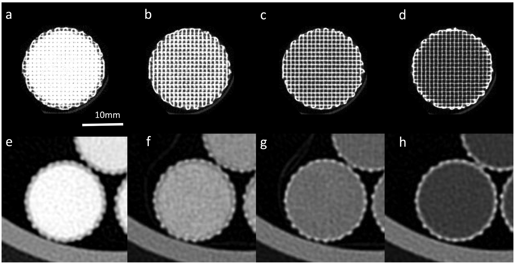
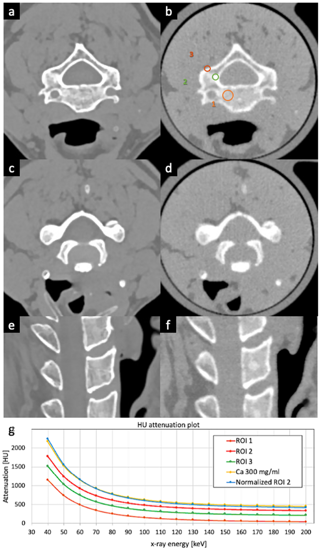
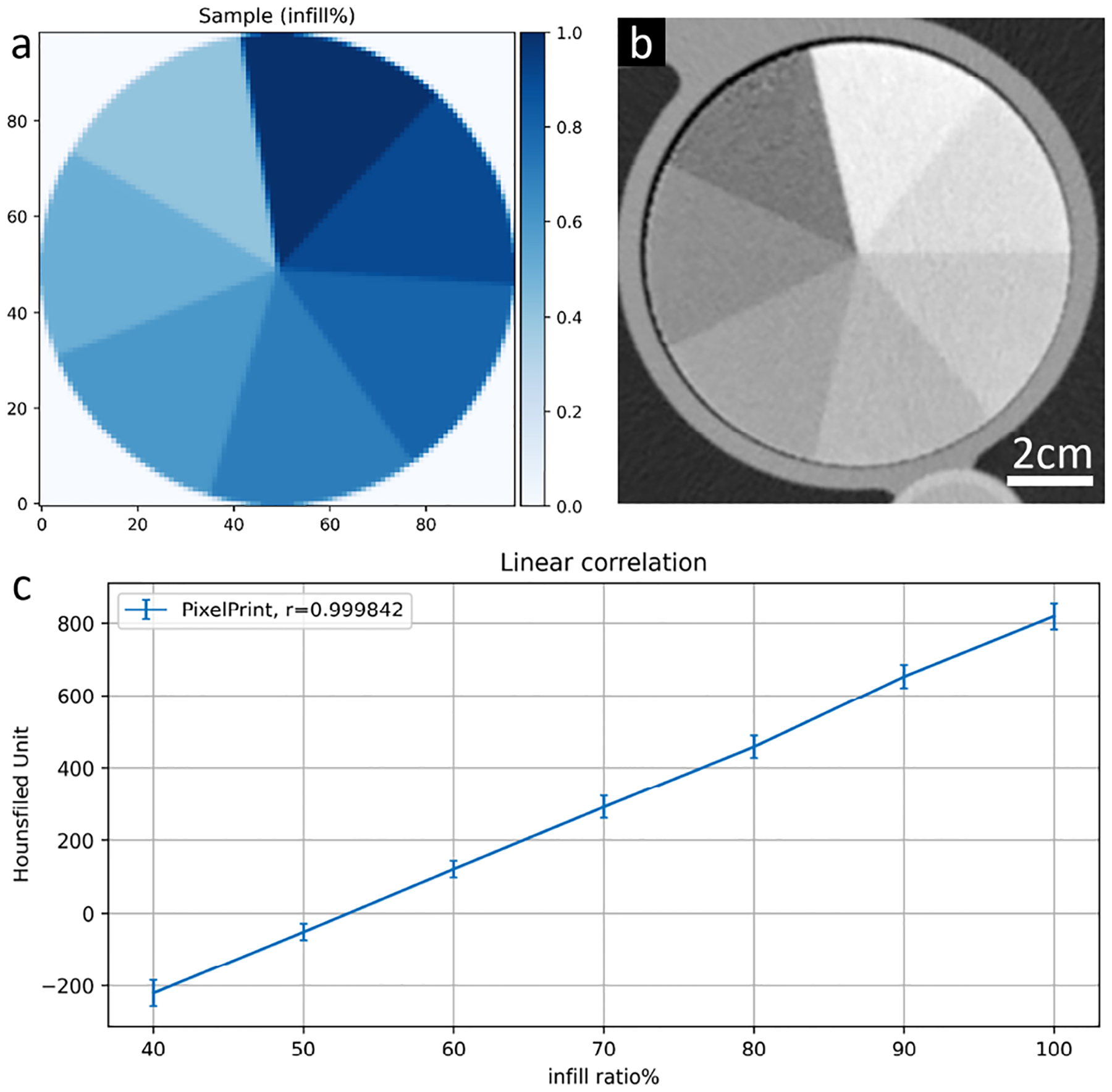
Patient-based CT phantoms, with realistic image texture and densities, are essential tools for assessing and verifying CT performance in clinical practice. This study extends our previously presented 3D printing solution (PixelPrint) to patient-based phantoms with soft tissue and bone structures. To expand the Hounsfield Unit (HUs) range, we utilize a stone-based filament. Applying PixelPrint, we converted patient DICOM images directly into FDM printer instructions (G-code). Density was modeled as the ratio of filament to voxel volume to emulate attenuation profiles for each voxel, with the filament ratio controlled through continuous modification of the printing speed. Two different phantoms were designed to demonstrate the high reproducibility of our approach with micro-CT acquisitions, and to determine the mapping between filament line widths and HU values on a clinical CT system. Moreover, a third phantom based on a clinical cervical spine scan was manufactured and scanned with a clinical spectral CT scanner. CT image of the patient-based phantom closely resembles the original CT image both in texture and contrast levels. Measured differences between patient and phantom are around 10 HU for bone marrow voxels and around 150 HU for cortical bone. In addition, stone-based filament can accurately represent boney tissue structures across the different x-ray energies, as measured by spectral CT. This study demonstrates the feasibility of our 3D-printed patient-based phantoms to be extended to soft-tissue and bone structure while maintaining accurate organ geometry, image texture, and attenuation profiles for spectral CT.
基于患者的CT体模具有逼真的图像纹理和密度,是临床实践中评估和验证CT性能的重要工具。本研究将我们之前提出的3D打印解决方案(PixelPrint)扩展到具有软组织和骨骼结构的基于患者的体模。为了扩大亨氏单位(HU)范围,我们使用了一种基于石头的细丝。应用PixelPrint,我们将患者的DICOM图像直接转换为FDM打印机指令(G代码)。密度被建模为细丝与体素体积的比率,以模拟每个体素的衰减曲线,细丝比率通过连续改变打印速度来控制。设计了两种不同的体模,以证明我们的方法在微CT采集中具有高再现性,并确定临床CT系统上细丝线宽与HU值之间的映射关系。此外,制造了一个基于临床颈椎扫描的第三个体模,并使用临床光谱CT扫描仪进行扫描。基于患者的体模CT图像在纹理和对比度水平上都与原始CT图像非常相似。对于骨髓体素,患者与体模之间的测量差异约为10 HU,对于皮质骨约为150 HU。此外,通过光谱CT测量,基于石头的细丝可以在不同的X射线能量下准确地呈现骨组织结构。本研究证明了我们3D打印的基于患者的体模扩展到软组织和骨骼结构的可行性,同时保持了光谱CT的准确器官几何形状、图像纹理和衰减曲线。