Department of Radiology, University of Pennsylvania, Philadelphia, PA, United States of America.
Department of Bioengineering, University of Pennsylvania, Philadelphia, PA, United States of America.
Phys Med Biol. 2024 May 14;69(11):115009. doi: 10.1088/1361-6560/ad3dba.
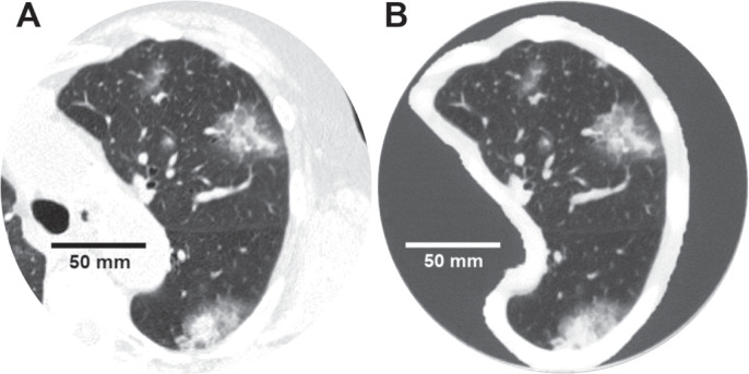
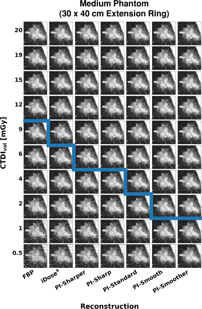
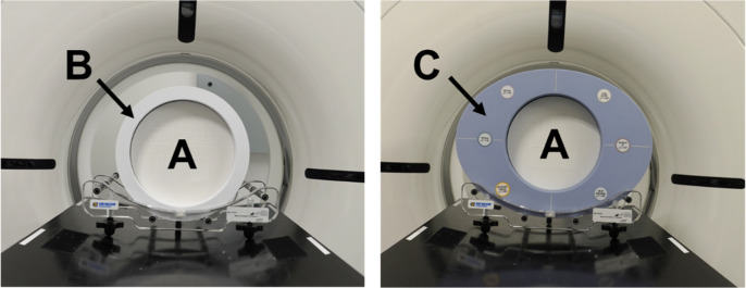
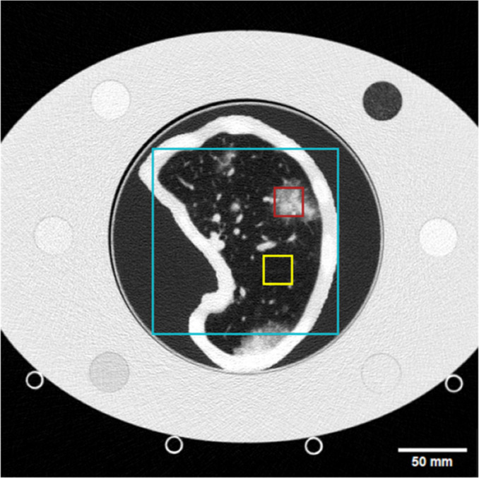
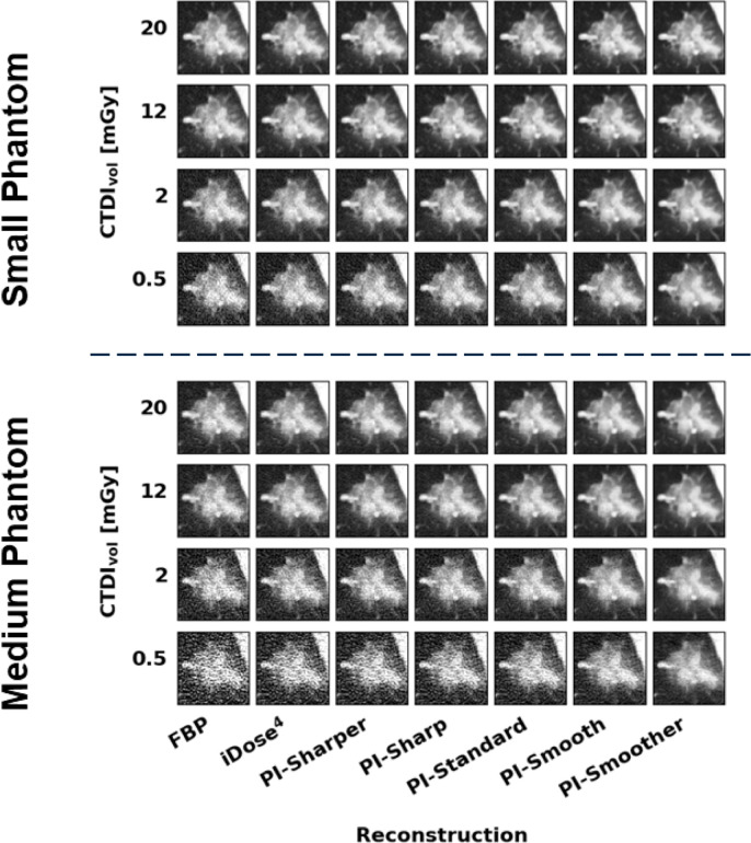
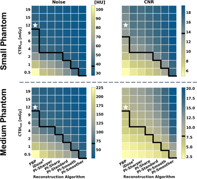
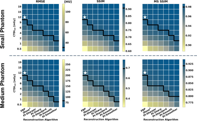
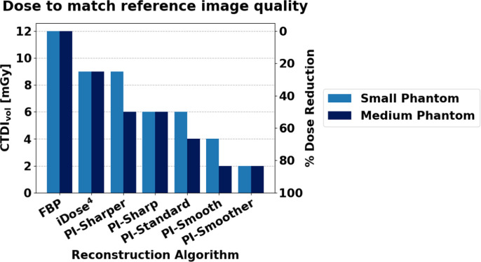
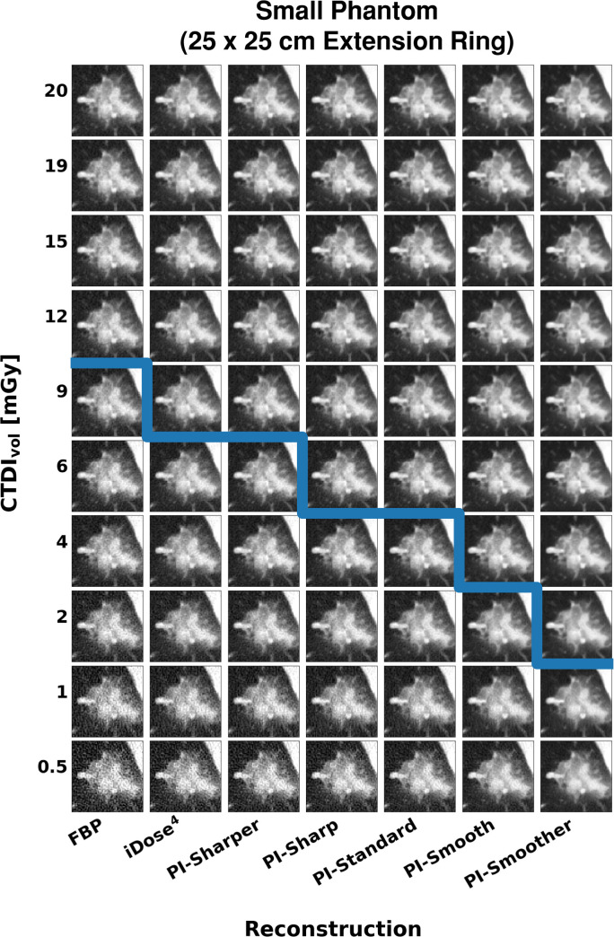
. Deep learning reconstruction (DLR) algorithms exhibit object-dependent resolution and noise performance. Thus, traditional geometric CT phantoms cannot fully capture the clinical imaging performance of DLR. This study uses a patient-derived 3D-printed PixelPrint lung phantom to evaluate a commercial DLR algorithm across a wide range of radiation dose levels.. The lung phantom used in this study is based on a patient chest CT scan containing ground glass opacities and was fabricated using PixelPrint 3D-printing technology. The phantom was placed inside two different size extension rings to mimic a small- and medium-sized patient and was scanned on a conventional CT scanner at exposures between 0.5 and 20 mGy. Each scan was reconstructed using filtered back projection (FBP), iterative reconstruction, and DLR at five levels of denoising. Image noise, contrast to noise ratio (CNR), root mean squared error, structural similarity index (SSIM), and multi-scale SSIM (MS SSIM) were calculated for each image.DLR demonstrated superior performance compared to FBP and iterative reconstruction for all measured metrics in both phantom sizes, with better performance for more aggressive denoising levels. DLR was estimated to reduce dose by 25%-83% in the small phantom and by 50%-83% in the medium phantom without decreasing image quality for any of the metrics measured in this study. These dose reduction estimates are more conservative compared to the estimates obtained when only considering noise and CNR.. DLR has the capability of producing diagnostic image quality at up to 83% lower radiation dose, which can improve the clinical utility and viability of lower dose CT scans. Furthermore, the PixelPrint phantom used in this study offers an improved testing environment with more realistic tissue structures compared to traditional CT phantoms, allowing for structure-based image quality evaluation beyond noise and contrast-based assessments.
深度学习重建(DLR)算法表现出与物体相关的分辨率和噪声性能。因此,传统的几何 CT 体模不能完全捕捉 DLR 的临床成像性能。本研究使用基于患者胸部 CT 扫描的 3D 打印 PixelPrint 肺体模来评估一种商业 DLR 算法在广泛的辐射剂量水平下的性能。本研究中使用的肺体模基于包含磨玻璃混浊的患者胸部 CT 扫描,并使用 PixelPrint 3D 打印技术制造。体模被放置在两个不同尺寸的延伸环内,以模拟小和中等体型的患者,并在常规 CT 扫描仪上以 0.5 至 20 mGy 的曝光量进行扫描。每个扫描均使用滤波反投影(FBP)、迭代重建和 DLR 在五个噪声水平进行重建。为每个图像计算图像噪声、对比噪声比(CNR)、均方根误差、结构相似性指数(SSIM)和多尺度 SSIM(MS SSIM)。在两种体模尺寸下,对于所有测量指标,DLR 均表现出优于 FBP 和迭代重建的性能,随着去噪水平的提高,性能也越好。对于本研究中测量的所有指标,DLR 估计可使小体模的剂量降低 25%至 83%,使中体模的剂量降低 50%至 83%,而不会降低图像质量。与仅考虑噪声和 CNR 时获得的估计相比,这些剂量减少估计更为保守。DLR 具有在低至 83%的辐射剂量下生成诊断图像质量的能力,这可以提高低剂量 CT 扫描的临床实用性和可行性。此外,与传统 CT 体模相比,本研究中使用的 PixelPrint 体模具有更真实的组织结构,提供了改进的测试环境,允许进行基于结构的图像质量评估,而不仅仅是基于噪声和对比度的评估。