• 文献检索
  • 文档翻译
  • 深度研究
  • 学术资讯
  • Suppr Zotero 插件Zotero 插件
  • 邀请有礼
  • 套餐&价格
  • 历史记录
应用&插件
Suppr Zotero 插件Zotero 插件浏览器插件Mac 客户端Windows 客户端微信小程序
定价
高级版会员购买积分包购买API积分包
服务
文献检索文档翻译深度研究API 文档MCP 服务
关于我们
关于 Suppr公司介绍联系我们用户协议隐私条款
关注我们

Suppr 超能文献

核心技术专利:CN118964589B侵权必究
粤ICP备2023148730 号-1Suppr @ 2026

文献检索

告别复杂PubMed语法,用中文像聊天一样搜索,搜遍4000万医学文献。AI智能推荐,让科研检索更轻松。

立即免费搜索

文件翻译

保留排版,准确专业,支持PDF/Word/PPT等文件格式,支持 12+语言互译。

免费翻译文档

深度研究

AI帮你快速写综述,25分钟生成高质量综述,智能提取关键信息,辅助科研写作。

立即免费体验

rrPDGF-BB诱导的大鼠骨髓间充质干细胞促进牵张成骨过程中的骨再生。

Rat bone marrow mesenchymal stem cells induced by rrPDGF-BB promotes bone regeneration during distraction osteogenesis.

作者信息

Wu Shuo, Zhang Lijie, Zhang Ruidan, Yang Kang, Wei Qin, Jia Qiyu, Guo Jian, Ma Chuang

机构信息

Department of Microrepair and Reconstruction, the First Affiliated Hospital of Xinjiang Medical University, Urumqi, China.

Department of Neurology, the Second Affiliated Hospital of Xinjiang Medical University, Urumqi, China.

出版信息

Front Bioeng Biotechnol. 2023 Mar 7;11:1110703. doi: 10.3389/fbioe.2023.1110703. eCollection 2023.

DOI:10.3389/fbioe.2023.1110703
PMID:36959901
原文链接:https://pmc.ncbi.nlm.nih.gov/articles/PMC10027703/
Abstract

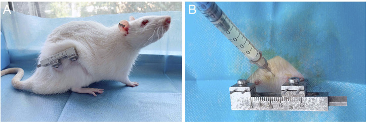
In the clinical treatment of large bone defects, distraction osteogenesis can be used. However, some patients may suffer from poor bone regeneration, or even delayed healing or non-union. Problems with the aggregation and proliferation of primary osteoblasts, or problems with the differentiation of primary osteoblasts will lead to poor bone regeneration. Therefore, supplementing exogenous primary osteoblasts and growth factors when using distraction osteogenesis may be a treatment plan with great potential. Bone marrow mesenchymal stem cells (BMSCs) were extracted from rats and cultured. Subsequently, Recombinant Rat Platelet-derived Growth Factor BB (rrPDGF-BB) was used to induce bone marrow mesenchymal stem cells. At the same time, male adult rats were selected to make the right femoral distraction osteogenesis model. During the mineralization period, phosphate buffer salt solution (control group), non-induction bone marrow mesenchymal stem cells (group 1) and recombinant rat platelet-derived growth factor BB intervened bone marrow mesenchymal stem cells (group 2) were injected into the distraction areas of each group. Then, the experimental results were evaluated with imaging and histology. Statistical analysis of the data showed that the difference was statistically significant if < 0.05. After intervention with recombinant rat platelet-derived growth factor BB on bone marrow mesenchymal stem cells, the cell morphology changed into a thin strip. After the cells were injected in the mineralization period, the samples showed that the callus in group 2 had greater hardness and the color close to the normal bone tissue; X-ray examination showed that there were more new callus in the distraction space of group 2; Micro-CT examination showed that there were more new bone tissues in group 2; Micro-CT data at week eight showed that the tissue volume, bone volume, percent bone volume, bone trabecular thickness, bone trabecular number and bone mineral density in group 2 were the largest, and the bone trabecular separation in group 2 was the smallest. There was a statistical difference between the groups ( < 0.05); HE staining confirmed that group 2 formed more blood vessels and chondrocytes earlier than the control group. At 8 weeks, the bone marrow cavity of group 2 was obvious, and some of them had been fused. The study confirmed that injecting bone marrow mesenchymal stem cellsBB into the distraction space of rats can promote the formation of new bone in the distraction area and promote the healing of distraction osteogenesis.

摘要

在大骨缺损的临床治疗中,可以采用牵张成骨术。然而,一些患者可能会出现骨再生不良的情况,甚至延迟愈合或不愈合。原代成骨细胞的聚集和增殖问题,或原代成骨细胞的分化问题会导致骨再生不良。因此,在使用牵张成骨术时补充外源性原代成骨细胞和生长因子可能是一个具有巨大潜力的治疗方案。从大鼠中提取骨髓间充质干细胞(BMSCs)并进行培养。随后,使用重组大鼠血小板衍生生长因子BB(rrPDGF-BB)诱导骨髓间充质干细胞。同时,选择成年雄性大鼠制作右侧股骨牵张成骨模型。在矿化期,将磷酸盐缓冲盐溶液(对照组)、未诱导的骨髓间充质干细胞(第1组)和重组大鼠血小板衍生生长因子BB干预的骨髓间充质干细胞(第2组)注入每组的牵张区域。然后,通过影像学和组织学对实验结果进行评估。数据的统计分析表明,如果<0.05,则差异具有统计学意义。用重组大鼠血小板衍生生长因子BB对骨髓间充质干细胞进行干预后,细胞形态变为细条状。在矿化期将细胞注射后,样本显示第2组的骨痂硬度更大,颜色接近正常骨组织;X线检查显示第2组牵张间隙中的新生骨痂更多;显微CT检查显示第2组的新骨组织更多;第8周的显微CT数据显示,第2组的组织体积、骨体积、骨体积百分比、骨小梁厚度、骨小梁数量和骨密度最大,第2组的骨小梁间距最小。各组之间存在统计学差异(<0.05);苏木精-伊红染色证实,第2组比对照组更早形成更多的血管和软骨细胞。在8周时,第2组的骨髓腔明显,其中一些已经融合。该研究证实,将骨髓间充质干细胞BB注入大鼠的牵张间隙可促进牵张区域新骨的形成,促进牵张成骨的愈合。

https://cdn.ncbi.nlm.nih.gov/pmc/blobs/8fdd/10027703/fd5b63c9d584/fbioe-11-1110703-g012.jpg
https://cdn.ncbi.nlm.nih.gov/pmc/blobs/8fdd/10027703/deff727cb774/fbioe-11-1110703-g001.jpg
https://cdn.ncbi.nlm.nih.gov/pmc/blobs/8fdd/10027703/8580707a349c/fbioe-11-1110703-g002.jpg
https://cdn.ncbi.nlm.nih.gov/pmc/blobs/8fdd/10027703/e464c8743146/fbioe-11-1110703-g003.jpg
https://cdn.ncbi.nlm.nih.gov/pmc/blobs/8fdd/10027703/9718f36985c3/fbioe-11-1110703-g004.jpg
https://cdn.ncbi.nlm.nih.gov/pmc/blobs/8fdd/10027703/a3df3b573b23/fbioe-11-1110703-g005.jpg
https://cdn.ncbi.nlm.nih.gov/pmc/blobs/8fdd/10027703/520168dcd9b7/fbioe-11-1110703-g006.jpg
https://cdn.ncbi.nlm.nih.gov/pmc/blobs/8fdd/10027703/dd50f94a021f/fbioe-11-1110703-g007.jpg
https://cdn.ncbi.nlm.nih.gov/pmc/blobs/8fdd/10027703/a9fc44488e0b/fbioe-11-1110703-g008.jpg
https://cdn.ncbi.nlm.nih.gov/pmc/blobs/8fdd/10027703/a45f410d898d/fbioe-11-1110703-g009.jpg
https://cdn.ncbi.nlm.nih.gov/pmc/blobs/8fdd/10027703/5fa7e6a51724/fbioe-11-1110703-g010.jpg
https://cdn.ncbi.nlm.nih.gov/pmc/blobs/8fdd/10027703/6efcd7751d3f/fbioe-11-1110703-g011.jpg
https://cdn.ncbi.nlm.nih.gov/pmc/blobs/8fdd/10027703/fd5b63c9d584/fbioe-11-1110703-g012.jpg

相似文献

1
Rat bone marrow mesenchymal stem cells induced by rrPDGF-BB promotes bone regeneration during distraction osteogenesis.rrPDGF-BB诱导的大鼠骨髓间充质干细胞促进牵张成骨过程中的骨再生。
Front Bioeng Biotechnol. 2023 Mar 7;11:1110703. doi: 10.3389/fbioe.2023.1110703. eCollection 2023.
2
Recombinant human platelet-derived growth factor-BB augmentation of new-bone formation in a rat model of distraction osteogenesis.重组人血小板衍生生长因子-BB促进大鼠牵张成骨模型中新生骨形成的研究
J Bone Joint Surg Am. 2009 Aug;91(8):1973-84. doi: 10.2106/JBJS.H.00540.
3
NELL1 promotes high-quality bone regeneration in rat femoral distraction osteogenesis model.NELL1 促进大鼠股骨牵张成骨模型中的高质量骨再生。
Bone. 2011 Mar 1;48(3):485-95. doi: 10.1016/j.bone.2010.10.166. Epub 2010 Oct 17.
4
Uncultured autogenous adipose-derived regenerative cells promote bone formation during distraction osteogenesis in rats.未培养的自体脂肪来源的再生细胞促进大鼠牵张成骨过程中的骨形成。
Clin Orthop Relat Res. 2014 Dec;472(12):3798-806. doi: 10.1007/s11999-014-3608-8.
5
Effects of bone morphogenetic protein 2 gene therapy on new bone formation during mandibular distraction osteogenesis at rapid rate in rabbits.骨形态发生蛋白2基因治疗对兔下颌快速牵张成骨过程中新骨形成的影响。
Oral Surg Oral Med Oral Pathol Oral Radiol Endod. 2011 Jul;112(1):50-7. doi: 10.1016/j.tripleo.2010.09.065. Epub 2010 Dec 30.
6
The effect of bone morphogenic protein-2-coated tri-calcium phosphate/hydroxyapatite on new bone formation in a rat model of femoral distraction osteogenesis.骨形成蛋白-2 涂层三钙磷/羟磷灰石对大鼠股骨牵张成骨中新骨形成的影响。
Cytotherapy. 2012 Mar;14(3):315-26. doi: 10.3109/14653249.2011.630728. Epub 2011 Nov 28.
7
Exosomes Secreted by Young Mesenchymal Stem Cells Promote New Bone Formation During Distraction Osteogenesis in Older Rats.年轻间充质干细胞分泌的外泌体在老年大鼠牵张成骨过程中促进新骨形成。
Calcif Tissue Int. 2020 May;106(5):509-517. doi: 10.1007/s00223-019-00656-4. Epub 2020 Feb 26.
8
PDGF-BB exhibited therapeutic effects on rat model of bisphosphonate-related osteonecrosis of the jaw by enhancing angiogenesis and osteogenesis.血小板衍生生长因子 BB 通过促进血管生成和骨生成对双膦酸盐相关性颌骨骨坏死大鼠模型发挥治疗作用。
Bone. 2021 Mar;144:115117. doi: 10.1016/j.bone.2019.115117. Epub 2019 Oct 30.
9
Transcription factor osterix modified bone marrow mesenchymal stem cells enhance callus formation during distraction osteogenesis.转录因子osterix修饰的骨髓间充质干细胞可增强牵张成骨过程中的骨痂形成。
Oral Surg Oral Med Oral Pathol Oral Radiol Endod. 2011 Apr;111(4):412-9. doi: 10.1016/j.tripleo.2010.05.012. Epub 2010 Sep 1.
10
[Effect of "accordion" technique on bone consolidation during distraction osteogenesis].["手风琴"技术对牵张成骨过程中骨愈合的影响]
Zhongguo Xiu Fu Chong Jian Wai Ke Za Zhi. 2018 May 15;32(5):558-567. doi: 10.7507/1002-1892.201712094.

引用本文的文献

1
Enabling adenosine signaling to promote aged fracture healing.激活腺苷信号以促进老年骨折愈合。
NPJ Regen Med. 2025 Apr 9;10(1):18. doi: 10.1038/s41536-025-00406-1.
2
Stem Cells: Present Understanding and Prospects for Regenerative Dentistry.干细胞:目前对再生牙科的认识与前景
J Funct Biomater. 2024 Oct 15;15(10):308. doi: 10.3390/jfb15100308.
3
Targeted Therapy of Acute Liver Injury via Cryptotanshinone-Loaded Biomimetic Nanoparticles Derived from Mesenchymal Stromal Cells Driven by Homing.通过归巢驱动的间充质基质细胞衍生的载有隐丹参酮的仿生纳米颗粒对急性肝损伤进行靶向治疗

本文引用的文献

1
The effect of different combinations of fixatives and decalcifying agents on rat and rabbit hard tissues, a guide for histologic processing.不同固定剂和脱钙剂组合对大鼠和兔硬组织的影响,组织学处理指南。
Acta Histochem. 2022 Dec;124(8):151962. doi: 10.1016/j.acthis.2022.151962. Epub 2022 Oct 10.
2
Hsp20 Promotes Endothelial Progenitor Cell Angiogenesis via Activation of PI3K/Akt Signaling Pathway under Hypoxia.热休克蛋白 20 通过激活低氧条件下的 PI3K/Akt 信号通路促进内皮祖细胞血管生成。
Tissue Eng Regen Med. 2022 Dec;19(6):1251-1266. doi: 10.1007/s13770-022-00481-1. Epub 2022 Aug 30.
3
Hypoxia During the Consolidation Phase of Distraction Osteogenesis Promotes Bone Regeneration.
Pharmaceutics. 2023 Dec 12;15(12):2764. doi: 10.3390/pharmaceutics15122764.
牵张成骨巩固期的低氧促进骨再生。
Front Physiol. 2022 Feb 22;13:804469. doi: 10.3389/fphys.2022.804469. eCollection 2022.
4
Platelet-derived growth factor-BB regenerates functional periodontal ligament in the tooth replantation.血小板衍生生长因子-BB 可在牙再植中再生功能性牙周韧带。
Sci Rep. 2022 Feb 25;12(1):3223. doi: 10.1038/s41598-022-06865-6.
5
Cyclic Distraction-Compression Dynamization Technique Enhances the Bone Formation During Distraction Osteogenesis.周期性牵张-压缩动力化技术可增强牵张成骨过程中的骨形成。
Front Bioeng Biotechnol. 2022 Jan 18;9:810723. doi: 10.3389/fbioe.2021.810723. eCollection 2021.
6
Strategies for large bone defect reconstruction after trauma, infections or tumour excision: a comprehensive review of the literature.创伤、感染或肿瘤切除后大骨缺损的重建策略:文献综述。
Eur J Med Res. 2021 Oct 2;26(1):118. doi: 10.1186/s40001-021-00593-9.
7
Optimization of decalcification techniques for histologic examination of the rat maxillary and mandibular incisors for toxicity studies.用于毒性研究的大鼠上颌和下颌切牙组织学检查的脱钙技术优化
J Histotechnol. 2022 Mar;45(1):2-9. doi: 10.1080/01478885.2021.1974780. Epub 2021 Sep 24.
8
Novel polychrome staining distinguishing osteochondral tissue and bone cells in decalcified paraffin sections.新型多色染色可区分脱钙石蜡切片中的软骨组织和骨细胞。
Cell Tissue Res. 2021 Sep;385(3):727-737. doi: 10.1007/s00441-021-03516-6. Epub 2021 Aug 19.
9
EGFL6 regulates angiogenesis and osteogenesis in distraction osteogenesis via Wnt/β-catenin signaling.EGFL6 通过 Wnt/β-catenin 信号通路调控牵张成骨中的血管生成和成骨。
Stem Cell Res Ther. 2021 Jul 22;12(1):415. doi: 10.1186/s13287-021-02487-3.
10
Fundamentals of bone vasculature: Specialization, interactions and functions.骨血管系统的基础:特化、相互作用及功能
Semin Cell Dev Biol. 2022 Mar;123:36-47. doi: 10.1016/j.semcdb.2021.06.025. Epub 2021 Jul 17.