Department of General Surgery, Zhongshan Hospital, Fudan University, 180 Fenglin Road, Shanghai, 200032, China.
Shanghai Clinical Nutrition Research Center, Shanghai, China.
BMC Cancer. 2023 Mar 28;23(1):279. doi: 10.1186/s12885-023-10736-2.
The purpose of this study is to explore the difference of abdominal fat and muscle composition, especially subcutaneous and visceral adipose tissue, in different stages of colorectal cancer (CRC).
Patients were divided into 4 groups: healthy controls (patients without colorectal polyp), polyp group (patients with colorectal polyp), cancer group (CRC patients without cachexia), and cachexia group (CRC patients with cachexia). Skeletal muscle (SM), subcutaneous adipose tissue (SAT), visceral adipose tissue (VAT), and intermuscular adipose tissue (IMAT) were assessed at the third lumbar level on computed tomography images obtained within 30 days before colonoscopy or surgery. One-way ANOVA and linear regression were used to analyze the difference of abdominal fat and muscle composition in different stages of CRC.
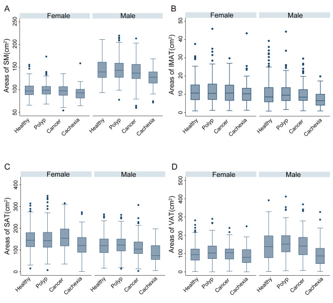
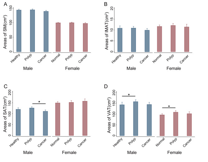
A total of 1513 patients were divided into healthy controls, polyp group, cancer group, and cachexia group, respectively. In the development of CRC from normal mucosa to polyp and cancer, the VAT area of the polyp group was significantly higher than that of the healthy controls both in male (156.32 ± 69.71 cm vs. 141.97 ± 79.40 cm, P = 0.014) and female patients (108.69 ± 53.95 cm vs. 96.28 ± 46.70 cm, P = 0.044). However, no significant differences were observed of SAT area between polyp group and healthy controls in both sexes. SAT area decreased significantly in the male cancer group compared with the polyp group (111.16 ± 46.98 cm vs. 126.40 ± 43.52 cm, P = 0.001), while no such change was observed in female patients. When compared with healthy controls, the SM, IMAT, SAT, and VAT areas of cachexia group was significantly decreased by 9.25 cm (95% CI: 5.39-13.11 cm, P < 0.001), 1.93 cm (95% CI: 0.54-3.32 cm, P = 0.001), 28.84 cm (95% CI: 17.84-39.83 cm, P < 0.001), and 31.31 cm (95% CI: 18.12-44.51 cm, P < 0.001) after adjusting for age and gender.
Abdominal fat and muscle composition, especially SAT and VAT, was differently distributed in different stages of CRC. It is necessary to pay attention to the different roles of subcutaneous and visceral adipose tissue in the development of CRC.
本研究旨在探讨结直肠癌(CRC)不同阶段腹部脂肪和肌肉成分的差异,尤其是皮下和内脏脂肪组织。
患者分为 4 组:健康对照组(无结直肠息肉患者)、息肉组(结直肠息肉患者)、癌症组(无恶液质的 CRC 患者)和恶液质组(有恶液质的 CRC 患者)。在结肠镜检查或手术前 30 天内获得的 CT 图像上,在第三腰椎水平评估骨骼肌(SM)、皮下脂肪组织(SAT)、内脏脂肪组织(VAT)和肌间脂肪组织(IMAT)。采用单因素方差分析和线性回归分析 CRC 不同阶段腹部脂肪和肌肉成分的差异。
共有 1513 名患者分为健康对照组、息肉组、癌症组和恶液质组。在从正常黏膜到息肉和癌症的 CRC 发展过程中,息肉组男性患者的 VAT 面积明显高于健康对照组(156.32 ± 69.71 cm 比 141.97 ± 79.40 cm,P = 0.014),女性患者也如此(108.69 ± 53.95 cm 比 96.28 ± 46.70 cm,P = 0.044)。然而,在两性中,息肉组与健康对照组的 SAT 面积无显著差异。与息肉组相比,男性癌症组的 SAT 面积显著下降(111.16 ± 46.98 cm 比 126.40 ± 43.52 cm,P = 0.001),而女性患者则无此变化。与健康对照组相比,恶液质组的 SM、IMAT、SAT 和 VAT 面积分别显著减少了 9.25 cm(95%CI:5.39-13.11 cm,P < 0.001)、1.93 cm(95%CI:0.54-3.32 cm,P = 0.001)、28.84 cm(95%CI:17.84-39.83 cm,P < 0.001)和 31.31 cm(95%CI:18.12-44.51 cm,P < 0.001),这些差异在调整年龄和性别后仍然存在。
CRC 不同阶段腹部脂肪和肌肉成分,尤其是 SAT 和 VAT 的分布不同。有必要关注皮下和内脏脂肪组织在 CRC 发展过程中的不同作用。