Suppr超能文献

深度学习方法在腹部 CT 定位与分割中的应用。

Deep learning method for localization and segmentation of abdominal CT.

机构信息

School of Engineering Science, Simon Fraser University, Canada.

School of Engineering Science, Simon Fraser University, Canada.

出版信息

Comput Med Imaging Graph. 2020 Oct;85:101776. doi: 10.1016/j.compmedimag.2020.101776. Epub 2020 Aug 14.

Abstract

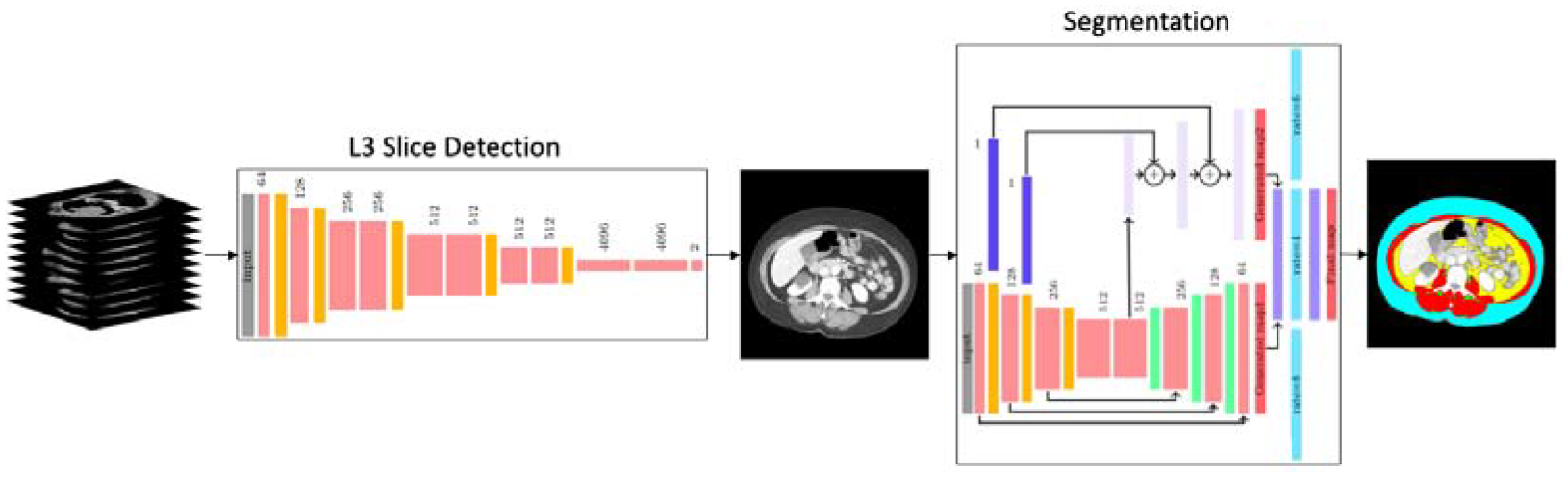
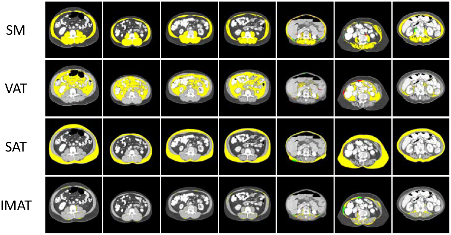
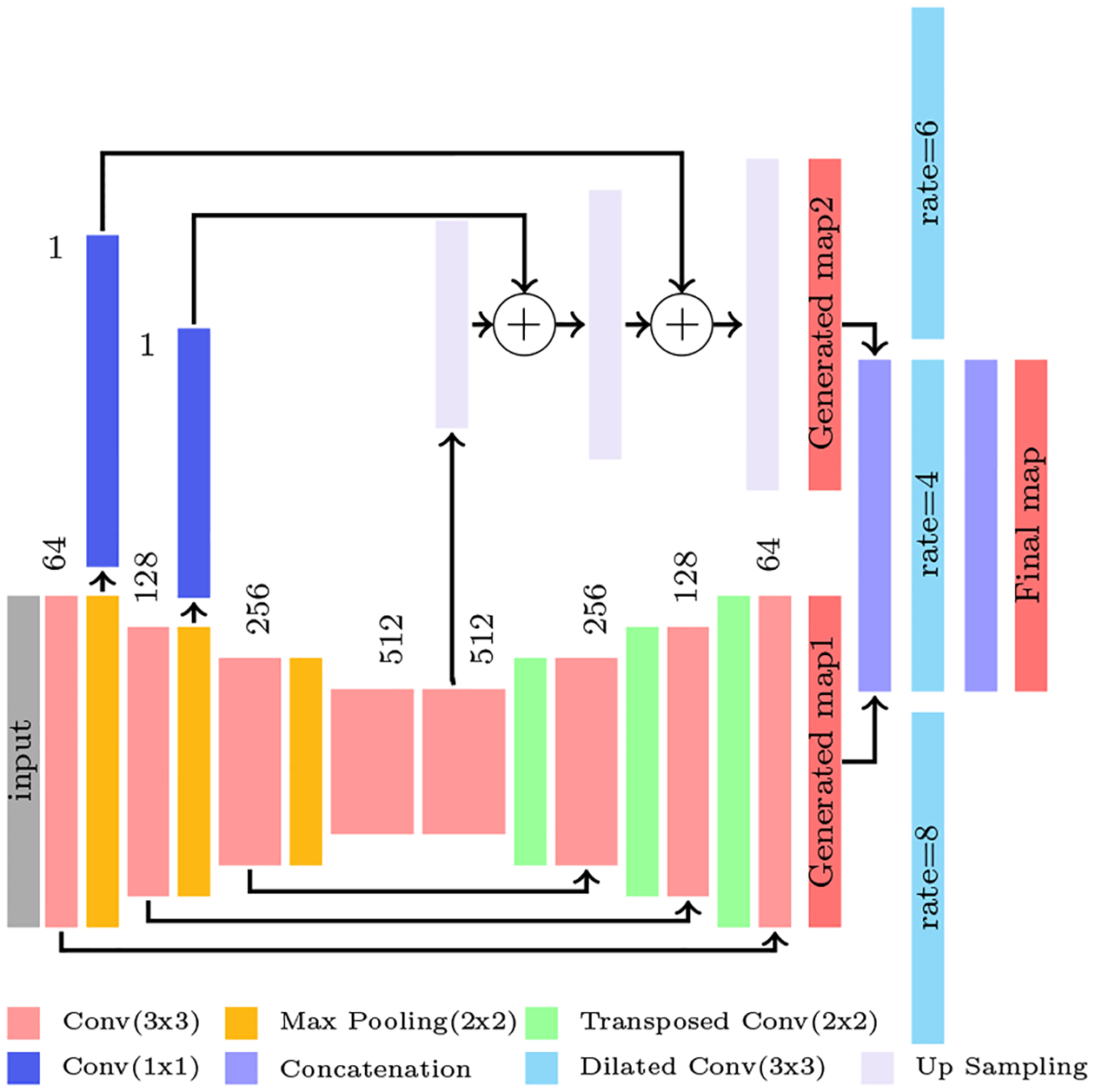
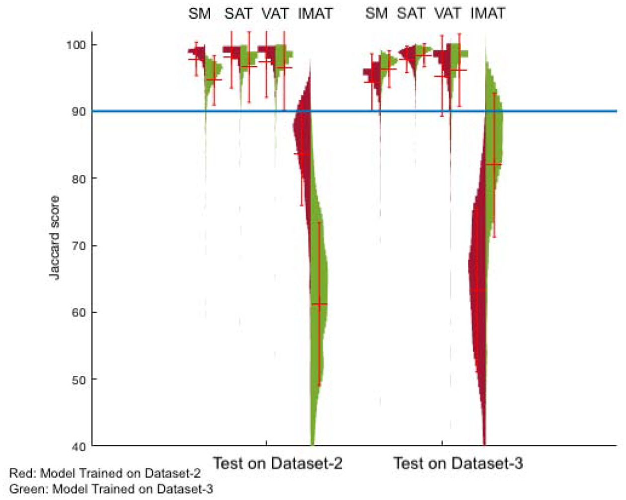
Computed Tomography (CT) imaging is widely used for studying body composition, i.e., the proportion of muscle and fat tissues with applications in areas such as nutrition or chemotherapy dose design. In particular, axial CT slices from the 3rd lumbar (L3) vertebral location are commonly used for body composition analysis. However, selection of the third lumbar vertebral slice and the segmentation of muscle/fat in the slice is a tedious operation if performed manually. The objective of this study is to automatically find the middle axial slice at L3 level from a full or partial body CT scan volume and segment the skeletal muscle (SM), subcutaneous adipose tissue (SAT), visceral adipose tissue (VAT) and intermuscular adipose tissue (IMAT) on that slice. The proposed algorithm includes an L3 axial slice localization network followed by a muscle-fat segmentation network. The localization network is a fully convolutional classifier trained on more than 12,000 images. The segmentation network is a convolutional neural network with an encoder-decoder architecture. Three datasets with CT images taken for patients with different types of cancers are used for training and validation of the networks. The mean slice error of 0.87±2.54 was achieved for L3 slice localization on 1748 CT scan volumes. The performance of five class tissue segmentation network evaluated on two datasets with 1327 and 1202 test samples. The mean Jaccard score of 97% was achieved for SM and VAT tissue segmentation on 1327 images. The mean Jaccard scores of 98% and 83% are corresponding to SAT and IMAT tissue segmentation on the same dataset. The localization and segmentation network performance indicates the potential for fully automated body composition analysis with high accuracy.

摘要

计算机断层扫描(CT)成像广泛用于研究身体成分,即肌肉和脂肪组织的比例,其应用领域包括营养或化疗剂量设计。特别是,从第三腰椎(L3)椎骨位置的轴向 CT 切片常用于身体成分分析。然而,如果手动执行,选择第三腰椎切片和对切片中的肌肉/脂肪进行分割是一项繁琐的操作。本研究的目的是从完整或部分身体 CT 扫描体积中自动找到 L3 水平的中间轴向切片,并对该切片上的骨骼肌(SM)、皮下脂肪组织(SAT)、内脏脂肪组织(VAT)和肌间脂肪组织(IMAT)进行分割。所提出的算法包括 L3 轴向切片定位网络和肌肉-脂肪分割网络。定位网络是一个基于超过 12000 张图像的全卷积分类器进行训练的。分割网络是一个具有编码器-解码器架构的卷积神经网络。该网络使用了三个包含不同类型癌症患者 CT 图像的数据集进行训练和验证。在 1748 个 CT 扫描体积上,L3 切片定位的平均切片误差为 0.87±2.54。在两个数据集上,使用 1327 个和 1202 个测试样本评估了五个组织分割网络的性能。在 1327 张图像上,SM 和 VAT 组织分割的平均 Jaccard 得分达到了 97%。在同一数据集上,SAT 和 IMAT 组织分割的平均 Jaccard 得分分别为 98%和 83%。定位和分割网络的性能表明,具有高精度的全自动身体成分分析具有潜力。

相似文献

1
Deep learning method for localization and segmentation of abdominal CT.
Comput Med Imaging Graph. 2020 Oct;85:101776. doi: 10.1016/j.compmedimag.2020.101776. Epub 2020 Aug 14.
4
Deep neural network for automatic volumetric segmentation of whole-body CT images for body composition assessment.
Clin Nutr. 2021 Aug;40(8):5038-5046. doi: 10.1016/j.clnu.2021.06.025. Epub 2021 Jul 15.
5
Automated segmentation of five different body tissues on computed tomography using deep learning.
Med Phys. 2023 Jan;50(1):178-191. doi: 10.1002/mp.15932. Epub 2022 Sep 2.
6
Body composition from single versus multi-slice abdominal computed tomography: Concordance and associations with colorectal cancer survival.
J Cachexia Sarcopenia Muscle. 2022 Dec;13(6):2974-2984. doi: 10.1002/jcsm.13080. Epub 2022 Sep 2.
8
Automatic segmentation of large-scale CT image datasets for detailed body composition analysis.
BMC Bioinformatics. 2023 Sep 18;24(1):346. doi: 10.1186/s12859-023-05462-2.
9
Deep learning for automated segmentation of pelvic muscles, fat, and bone from CT studies for body composition assessment.
Skeletal Radiol. 2020 Mar;49(3):387-395. doi: 10.1007/s00256-019-03289-8. Epub 2019 Aug 8.
10
A two-step convolutional neural network based computer-aided detection scheme for automatically segmenting adipose tissue volume depicting on CT images.
Comput Methods Programs Biomed. 2017 Jun;144:97-104. doi: 10.1016/j.cmpb.2017.03.017. Epub 2017 Mar 21.

引用本文的文献

1
Deep Learning-Based 3D and 2D Approaches for Skeletal Muscle Segmentation on Low-Dose CT Images.
J Imaging Inform Med. 2025 Aug 27. doi: 10.1007/s10278-025-01646-9.
2
Reliable Radiologic Skeletal Muscle Area Assessment - A Biomarker for Cancer Cachexia Diagnosis.
medRxiv. 2025 Apr 25:2025.04.21.25326162. doi: 10.1101/2025.04.21.25326162.
4
Body composition metrics as a determinant of trastuzumab deruxtecan related toxicity and response.
NPJ Breast Cancer. 2025 Apr 25;11(1):38. doi: 10.1038/s41523-025-00754-7.
5
Evolving perspectives on evaluating obesity: from traditional methods to cutting-edge techniques.
Ann Med. 2025 Dec;57(1):2472856. doi: 10.1080/07853890.2025.2472856. Epub 2025 Mar 12.
6

本文引用的文献

1
Muscle segmentation in axial computed tomography (CT) images at the lumbar (L3) and thoracic (T4) levels for body composition analysis.
Comput Med Imaging Graph. 2019 Jul;75:47-55. doi: 10.1016/j.compmedimag.2019.04.007. Epub 2019 May 9.
4
Spotting L3 slice in CT scans using deep convolutional network and transfer learning.
Comput Biol Med. 2017 Aug 1;87:95-103. doi: 10.1016/j.compbiomed.2017.05.018. Epub 2017 May 19.
5
Explaining the Obesity Paradox: The Association between Body Composition and Colorectal Cancer Survival (C-SCANS Study).
Cancer Epidemiol Biomarkers Prev. 2017 Jul;26(7):1008-1015. doi: 10.1158/1055-9965.EPI-17-0200. Epub 2017 May 15.
6
Fully Convolutional Networks for Semantic Segmentation.
IEEE Trans Pattern Anal Mach Intell. 2017 Apr;39(4):640-651. doi: 10.1109/TPAMI.2016.2572683. Epub 2016 May 24.
7
Body Composition Assessment in Axial CT Images Using FEM-Based Automatic Segmentation of Skeletal Muscle.
IEEE Trans Med Imaging. 2016 Feb;35(2):512-20. doi: 10.1109/TMI.2015.2479252. Epub 2015 Sep 22.
9
Skeletal muscle atrophy and increased intramuscular fat after incomplete spinal cord injury.
Spinal Cord. 2007 Apr;45(4):304-9. doi: 10.1038/sj.sc.3101968. Epub 2006 Aug 29.
10
Total body skeletal muscle and adipose tissue volumes: estimation from a single abdominal cross-sectional image.
J Appl Physiol (1985). 2004 Dec;97(6):2333-8. doi: 10.1152/japplphysiol.00744.2004. Epub 2004 Aug 13.

文献AI研究员

20分钟写一篇综述,助力文献阅读效率提升50倍。

立即体验

用中文搜PubMed

大模型驱动的PubMed中文搜索引擎

马上搜索

文档翻译

学术文献翻译模型,支持多种主流文档格式。

立即体验