Department of Radiology, University of Iowa, 200 Hawkins Drive cc704 GH, Iowa City, IA, 52242, USA.
Department of Biomedical Engineering, University of Iowa, Iowa City, IA, USA.
Sci Rep. 2023 Mar 29;13(1):5146. doi: 10.1038/s41598-023-32071-z.
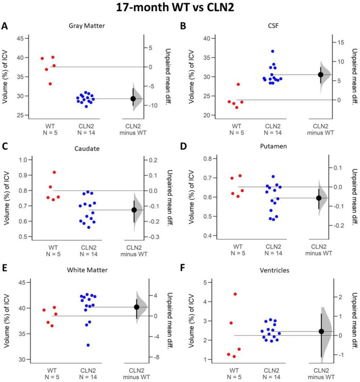
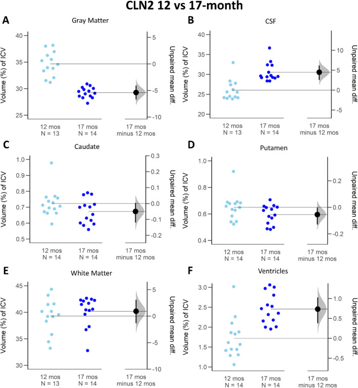
Late-infantile neuronal ceroid lipofuscinosis type 2 (CLN2) disease (Batten disease) is a rare pediatric disease, with symptom development leading to clinical diagnosis. Early diagnosis and effective tracking of disease progression are required for treatment. We hypothesize that brain volumetry is valuable in identifying CLN2 disease at an early stage and tracking disease progression in a genetically modified miniswine model. CLN2 miniswine and wild type controls were evaluated at 12- and 17-months of age, correlating to early and late stages of disease progression. Magnetic resonance imaging (MRI) T1- and T2-weighted data were acquired. Total intercranial, gray matter, cerebrospinal fluid, white matter, caudate, putamen, and ventricle volumes were calculated and expressed as proportions of the intracranial volume. The brain regions were compared between timepoints and cohorts using Gardner-Altman plots, mean differences, and confidence intervals. At an early stage of disease, the total intracranial volume (- 9.06 cm), gray matter (- 4.37% 95 CI - 7.41; - 1.83), caudate (- 0.16%, 95 CI - 0.24; - 0.08) and putamen (- 0.11% 95 CI - 0.23; - 0.02) were all notably smaller in CLN2 miniswines versus WT, while cerebrospinal fluid was larger (+ 3.42%, 95 CI 2.54; 6.18). As the disease progressed to a later stage, the difference between the gray matter (- 8.27%, 95 CI - 10.1; - 5.56) and cerebrospinal fluid (+ 6.88%, 95 CI 4.31; 8.51) continued to become more pronounced, while others remained stable. MRI brain volumetry in this miniswine model of CLN2 disease is sensitive to early disease detection and longitudinal change monitoring, providing a valuable tool for pre-clinical treatment development and evaluation.
晚婴型神经元蜡样脂褐质沉积症 2 型(CLN2)病(Batten 病)是一种罕见的儿科疾病,其症状发展导致临床诊断。需要早期诊断和有效跟踪疾病进展以进行治疗。我们假设脑容积测量在早期识别 CLN2 病和跟踪遗传修饰小型猪模型中的疾病进展方面具有价值。在 12 个月和 17 个月大时评估 CLN2 小型猪和野生型对照,分别对应疾病进展的早期和晚期。采集磁共振成像(MRI)T1-和 T2 加权数据。计算总颅内、灰质、脑脊液、白质、尾状核、壳核和脑室体积,并表示为颅内体积的比例。使用 Gardner-Altman 图、均值差异和置信区间比较脑区在时间点和队列之间的差异。在疾病的早期阶段,总颅内体积(-9.06cm)、灰质(-4.37%,95%置信区间-7.41;-1.83)、尾状核(-0.16%,95%置信区间-0.24;-0.08)和壳核(-0.11%,95%置信区间-0.23;-0.02)在 CLN2 小型猪中均显著小于 WT,而脑脊液更大(+3.42%,95%置信区间 2.54;6.18)。随着疾病进展到晚期,灰质(-8.27%,95%置信区间-10.1;-5.56)和脑脊液(+6.88%,95%置信区间 4.31;8.51)之间的差异继续变得更加明显,而其他差异则保持稳定。CLN2 病小型猪模型中的 MRI 脑容积测量对早期疾病检测和纵向变化监测敏感,为临床前治疗开发和评估提供了有价值的工具。