Barrow Rhiannon, Wilkinson Joseph N, He Yichen, Callaghan Martin, Brüning-Richardson Anke, Dunning Mark, Stead Lucy F
Leeds Institute of Medical Research at St James's, Wellcome Trust Brenner Building, St James's University Hospital, Leeds LS9 7TF, West Yorkshire, UK.
Department of Animal and Plant Sciences, Alfred Denny Building, The University of Sheffield, Western Bank, Sheffield S10 2TN, UK.
J Biol Methods. 2022 Nov 23;9(4):e163. doi: 10.14440/jbm.2022.388. eCollection 2022.
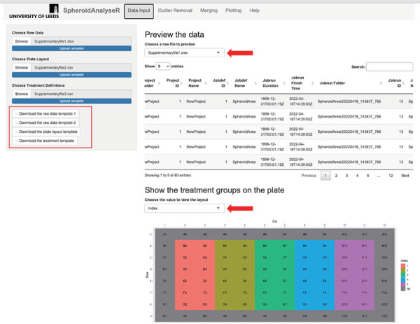
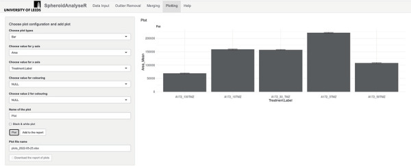
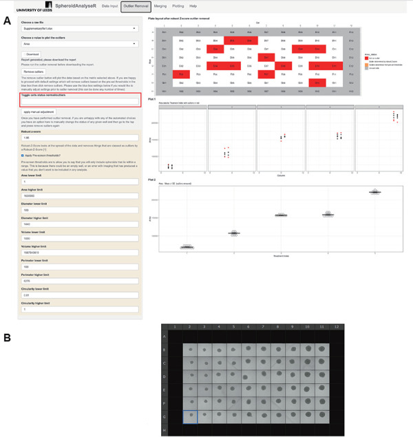
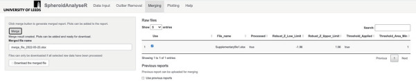
Spheroids and organoids are increasingly popular three-dimensional (3D) cell culture models. Spheroid models are more physiologically relevant to a tumor compared to two-dimensional (2D) cultures and organoids are a simplified version of an organ with similar composition. Spheroids are often only formed from a single cell type which does not represent the situation . However, despite this, both spheroids and organoids can be used in cell migration studies, disease modelling and drug discovery. A drawback of these models is, however, the lack of appropriate analytical tools for high throughput imaging and analysis over a time course. To address this, we have developed an R Shiny app called SpheroidAnalyseR: a simple, fast, effective open-source app that allows the analysis of spheroid or organoid size data generated in a 96-well format. SpheroidAnalyseR processes and analyzes datasets of image measurements that can be obtained a bespoke software, described herein, that automates spheroid imaging and quantification using the Nikon A1R Confocal Laser Scanning Microscope. However, templates are provided to enable users to input spheroid image measurements obtained by user-preferred methods. SpheroidAnalyseR facilitates outlier identification and removal followed by graphical visualization of spheroid measurements across multiple predefined parameters such as time, cell-type and treatment(s). Spheroid imaging and analysis can, thus, be reduced from hours to minutes, removing the requirement for substantial manual data manipulation in a spreadsheet application. The combination of spheroid generation in 96-well ultra-low attachment microplates, imaging using our bespoke software, and analysis using SpheroidAnalyseR toolkit allows high throughput, longitudinal quantification of 3D spheroid growth whilst minimizing user input and significantly improving the efficiency and reproducibility of data analysis. Our bespoke imaging software is available from https://github.com/GliomaGenomics. SpheroidAnalyseR is available at https://spheroidanalyser.leeds.ac.uk, and the source code found at https://github.com/GliomaGenomics.
球体和类器官是越来越受欢迎的三维(3D)细胞培养模型。与二维(2D)培养相比,球体模型在生理上与肿瘤更相关,而类器官是具有相似组成的器官的简化版本。球体通常仅由单一细胞类型形成,这并不代表实际情况。然而,尽管如此,球体和类器官都可用于细胞迁移研究、疾病建模和药物发现。然而,这些模型的一个缺点是缺乏适用于高通量成像和随时间过程分析的分析工具。为了解决这个问题,我们开发了一个名为SpheroidAnalyseR的R Shiny应用程序:一个简单、快速、有效的开源应用程序,可用于分析以96孔格式生成的球体或类器官大小数据。SpheroidAnalyseR处理和分析图像测量数据集,这些数据集可通过本文所述的定制软件获得,该软件使用尼康A1R共聚焦激光扫描显微镜自动进行球体成像和定量。不过,也提供了模板,以便用户输入通过其首选方法获得的球体图像测量数据。SpheroidAnalyseR有助于识别和去除异常值,然后对多个预定义参数(如时间、细胞类型和处理方式)的球体测量数据进行图形可视化。因此,球体成像和分析时间可从数小时缩短至数分钟,无需在电子表格应用程序中进行大量手动数据处理。在96孔超低附着微孔板中生成球体、使用我们的定制软件进行成像以及使用SpheroidAnalyseR工具包进行分析,这三者的结合可实现对3D球体生长的高通量纵向定量,同时将用户输入降至最低,并显著提高数据分析的效率和可重复性。我们的定制成像软件可从https://github.com/GliomaGenomics获取。SpheroidAnalyseR可在https://spheroidanalyser.leeds.ac.uk获取,其源代码可在https://github.com/GliomaGenomics找到。