Sakhy Youssef, Taoussi Reda, Ndayishimiye Vianney, Sabiri Mouna, Labied Mohammed, Lembarki Ghizlane, El Manjra Samia, Lezar Samira, Essodegui Fatiha
Central Radiology Department, Ibn Rochd University Hospital, Casablanca, Morocco.
Emergency Radiology Department, Ibn Rochd University Hospital, Casablanca, Morocco.
BJR Case Rep. 2023 Feb 15;9(2):20220083. doi: 10.1259/bjrcr.20220083. eCollection 2023 Mar.
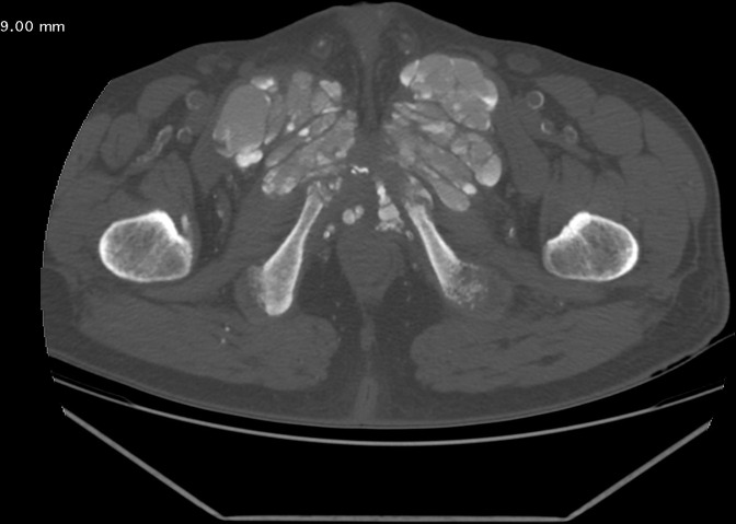
Tumoral calcinosis is a rare cause of intratissular calcifications in hemodialysis patients with chronic renal failure. Its frequency is estimated between 0.5 and 7% of patients. We illustrate through a case of unusual localization diagnosed in Ibn Rochd University Hospital, Casablanca, Morocco, the radiographic and scannographic aspect of this little known entity. A 40-year-old man, followed for hypertensive cardiopathy, in chronic renal failure for 12 years under hemodialysis, consulted for bilateral inguinal swellings evolving in a progressive and painless way. Biological investigations revealed hyperparathyroidism with increased phosphocalcic product. He was referred to us for radiological evaluation which revealed lesions in favor of bilateral puboinguinal tumor calcinosis. Tumoral calcinosis is a rare cause of intratissular calcifications in chronic renal failure patients undergoing hemodialysis. Pubic localization with infiltration and osteolysis of the symphysis pubis is very rare. Its main risk factors are the existence of hyperparathyroidism, an increase in phosphocalcic product and probably local traumatic factors. Tumoral calcinosis has a typical appearance on radiographs: amorphous, cystic and multilobulated calcifications of periarticular distribution. The CT scan allows a better delineation of the calcified mass. Its treatment remains controversial. The knowledge of osteoarticular manifestations of chronic hemodialysis patients, especially tumoral calcinosis by radiologists, allows to easily make the diagnosis and thus avoid invasive complementary explorations for the patient and to quickly institute an effective treatment.
肿瘤性钙化是慢性肾衰竭血液透析患者组织内钙化的罕见原因。其发生率估计在患者总数的0.5%至7%之间。我们通过在摩洛哥卡萨布兰卡伊本·鲁世德大学医院诊断出的一例罕见部位的病例,阐述了这个鲜为人知的病症的影像学表现。一名40岁男性,因高血压性心脏病接受治疗,已慢性肾衰竭12年,一直在进行血液透析,因双侧腹股沟逐渐无痛性肿大前来就诊。实验室检查显示甲状旁腺功能亢进,钙磷乘积升高。他被转诊至我们科室进行放射学评估,结果显示病变支持双侧耻骨腹股沟肿瘤性钙化。肿瘤性钙化是接受血液透析的慢性肾衰竭患者组织内钙化的罕见原因。耻骨部位伴有耻骨联合浸润和骨质溶解的情况非常罕见。其主要危险因素包括甲状旁腺功能亢进、钙磷乘积升高以及可能的局部创伤因素。肿瘤性钙化在X线片上有典型表现:关节周围分布的无定形、囊性和多叶状钙化。CT扫描能更好地勾勒出钙化肿块。其治疗仍存在争议。放射科医生了解慢性血液透析患者的骨关节表现,尤其是肿瘤性钙化,有助于轻松做出诊断,从而避免患者进行侵入性的辅助检查,并能迅速开展有效的治疗。