Laboratory of Clinical Neurochemistry, Section of Neurology, Department of Medicine and Surgery, University of Perugia, Piazzale Lucio Severi 1/8, 06132, Perugia, Italy.
R&D Unit, Amprion Inc, 11095 Flintkote Av., San Diego, San Diego, CA, 92121, USA.
Mol Neurodegener. 2023 Apr 1;18(1):20. doi: 10.1186/s13024-023-00613-8.
Aggregation of α-synuclein (α-syn) is a prominent feature of Parkinson's disease (PD) and other synucleinopathies. Currently, α-syn seed amplification assays (SAAs) using cerebrospinal fluid (CSF) represent the most promising diagnostic tools for synucleinopathies. However, CSF itself contains several compounds that can modulate the aggregation of α-syn in a patient-dependent manner, potentially undermining unoptimized α-syn SAAs and preventing seed quantification.
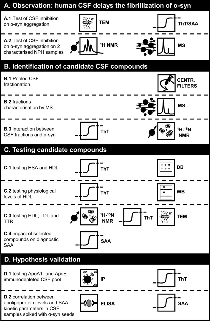
In this study, we characterized the inhibitory effect of CSF milieu on detection of α-syn aggregates by means of CSF fractionation, mass spectrometry, immunoassays, transmission electron microscopy, solution nuclear magnetic resonance spectroscopy, a highly accurate and standardized diagnostic SAA, and different in vitro aggregation conditions to evaluate spontaneous aggregation of α-syn.
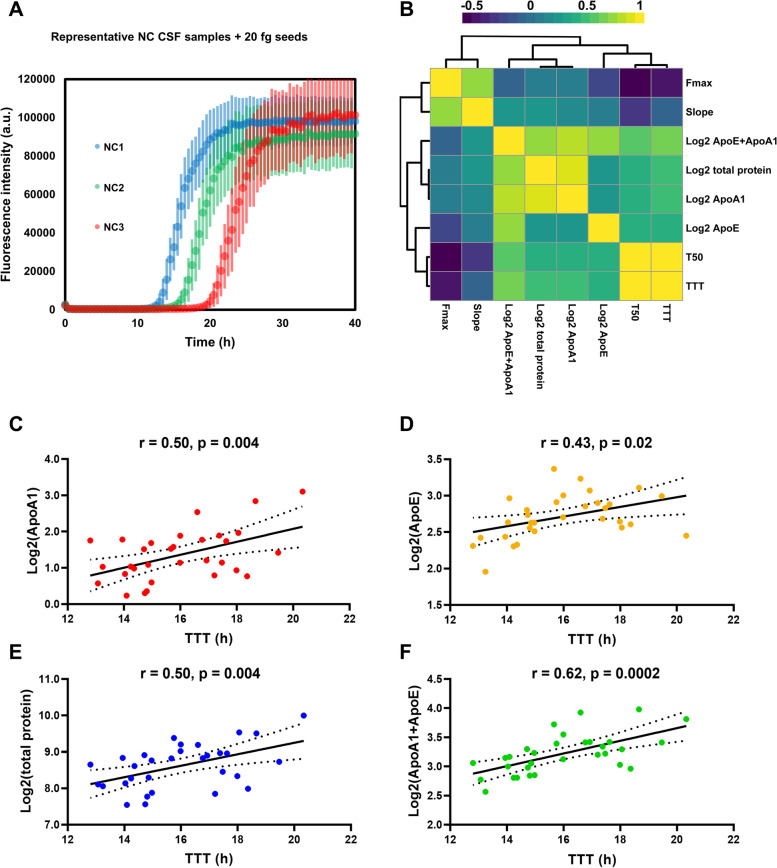
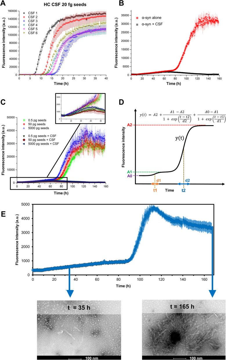
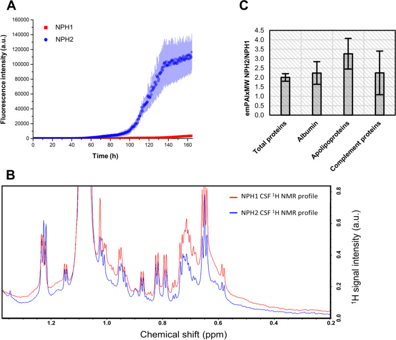
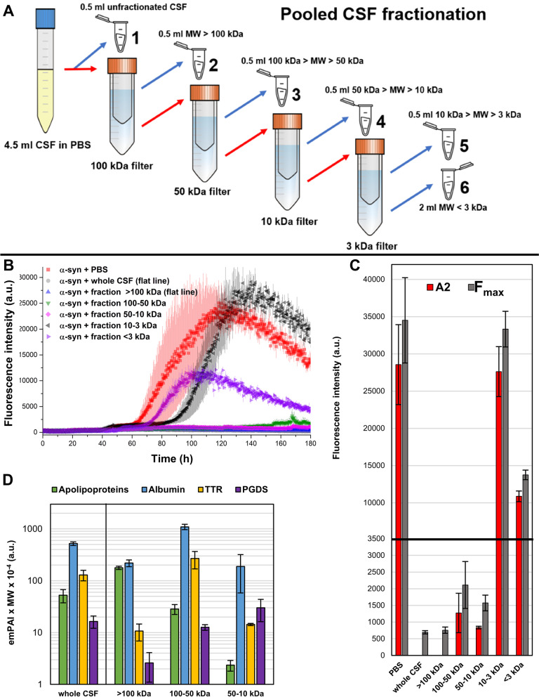
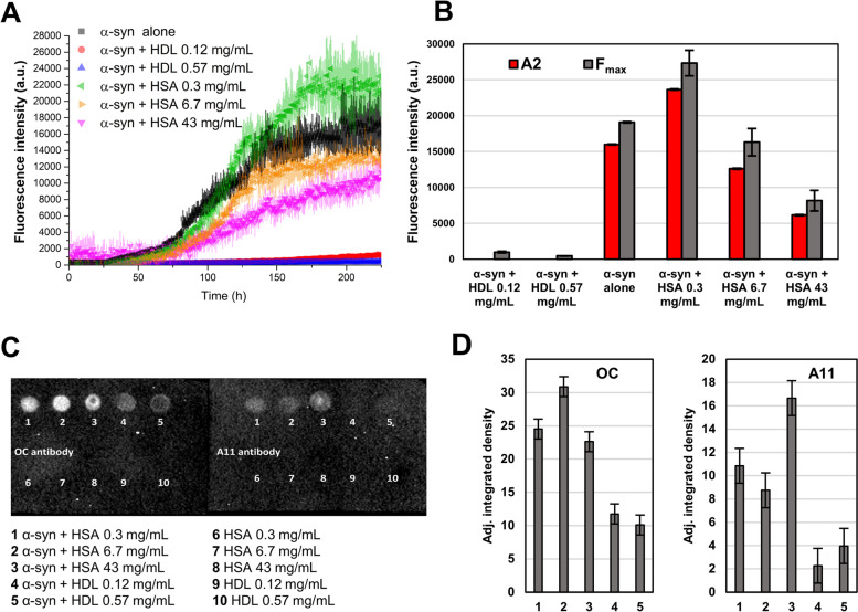
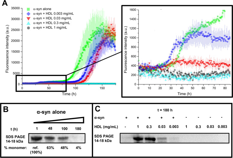
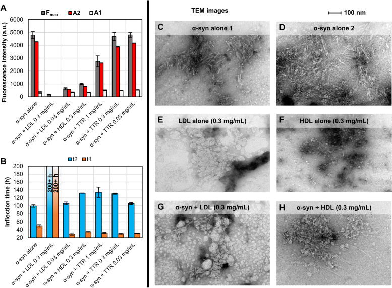
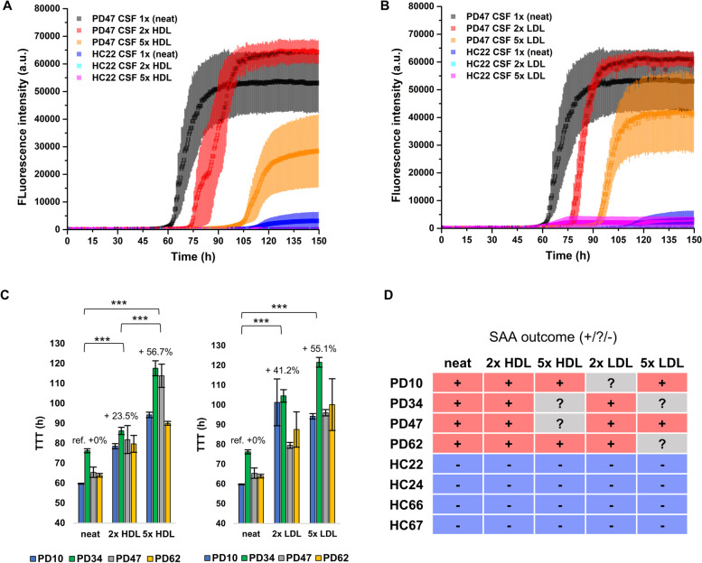
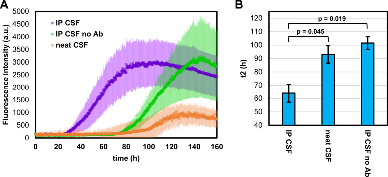
We found the high-molecular weight fraction of CSF (> 100,000 Da) to be highly inhibitory on α-syn aggregation and identified lipoproteins to be the main drivers of this effect. Direct interaction between lipoproteins and monomeric α-syn was not detected by solution nuclear magnetic resonance spectroscopy, on the other hand we observed lipoprotein-α-syn complexes by transmission electron microscopy. These observations are compatible with hypothesizing an interaction between lipoproteins and oligomeric/proto-fibrillary α-syn intermediates. We observed significantly slower amplification of α-syn seeds in PD CSF when lipoproteins were added to the reaction mix of diagnostic SAA. Additionally, we observed a decreased inhibition capacity of CSF on α-syn aggregation after immunodepleting ApoA1 and ApoE. Finally, we observed that CSF ApoA1 and ApoE levels significantly correlated with SAA kinetic parameters in n = 31 SAA-negative control CSF samples spiked with preformed α-syn aggregates.
Our results describe a novel interaction between lipoproteins and α-syn aggregates that inhibits the formation of α-syn fibrils and could have relevant implications. Indeed, the donor-specific inhibition of CSF on α-syn aggregation explains the lack of quantitative results from analysis of SAA-derived kinetic parameters to date. Furthermore, our data show that lipoproteins are the main inhibitory components of CSF, suggesting that lipoprotein concentration measurements could be incorporated into data analysis models to eliminate the confounding effects of CSF milieu on α-syn quantification efforts.
α-突触核蛋白(α-syn)的聚集是帕金森病(PD)和其他突触核蛋白病的一个显著特征。目前,使用脑脊液(CSF)的α-syn 种子扩增测定法(SAA)代表了突触核蛋白病最有前途的诊断工具。然而,CSF 本身包含几种化合物,这些化合物可以以患者依赖的方式调节α-syn 的聚集,从而破坏未优化的α-syn SAA,并防止种子定量。
在这项研究中,我们通过 CSF 分级分离、质谱、免疫测定、透射电子显微镜、溶液核磁共振光谱、高度准确和标准化的诊断 SAA 以及不同的体外聚合条件来表征 CSF 环境对检测α-syn 聚集物的抑制作用,以评估α-syn 的自发聚合。
我们发现 CSF 的高分子量部分(>100,000 Da)对α-syn 聚集具有高度抑制作用,并确定脂蛋白是这种作用的主要驱动因素。另一方面,通过溶液核磁共振光谱未检测到脂蛋白与单体α-syn 之间的直接相互作用,我们通过透射电子显微镜观察到脂蛋白-α-syn 复合物。这些观察结果与脂蛋白与寡聚体/原纤维状α-syn 中间体之间的相互作用假说一致。我们发现,当诊断 SAA 的反应混合物中添加脂蛋白时,PD CSF 中α-syn 种子的扩增明显较慢。此外,在免疫耗尽 ApoA1 和 ApoE 后,我们观察到 CSF 对α-syn 聚集的抑制能力降低。最后,我们观察到 CSF ApoA1 和 ApoE 水平与 n=31 个 SAA 阴性对照 CSF 样品中预形成的α-syn 聚集体的 SAA 动力学参数显著相关。
我们的结果描述了脂蛋白与α-syn 聚集体之间的一种新的相互作用,这种相互作用抑制了α-syn 纤维的形成,可能具有相关意义。事实上,CSF 对α-syn 聚集的供体特异性抑制解释了迄今为止分析 SAA 衍生动力学参数时缺乏定量结果的原因。此外,我们的数据表明,脂蛋白是 CSF 的主要抑制成分,这表明脂蛋白浓度测量可以纳入数据分析模型,以消除 CSF 环境对α-syn 定量工作的干扰。