Mast Cell Biology Section, Laboratory of Allergic Diseases, National Institute of Allergy and Infectious Diseases, National Institutes of Health, Bethesda, MD, United States.
National Institute of Allergy and Infectious Diseases (NIAID), Collaborative Bioinformatics Resource (NCBR), National Institute of Allergy and Infectious Diseases, National Institutes of Health, Bethesda, MD, United States.
Front Immunol. 2023 Mar 21;14:1078958. doi: 10.3389/fimmu.2023.1078958. eCollection 2023.
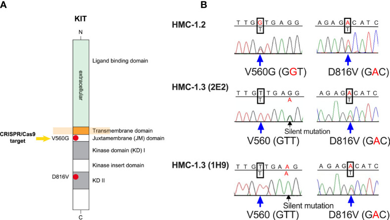
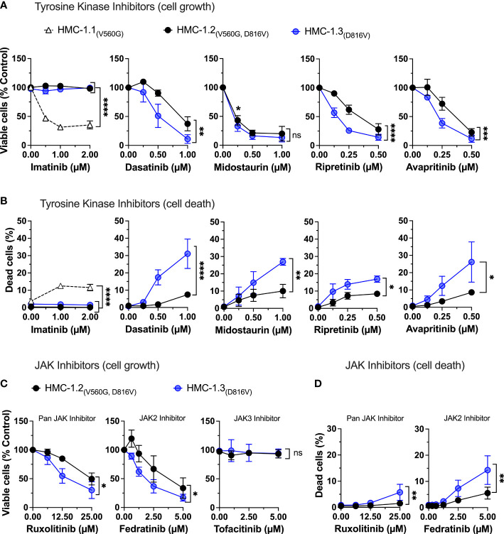
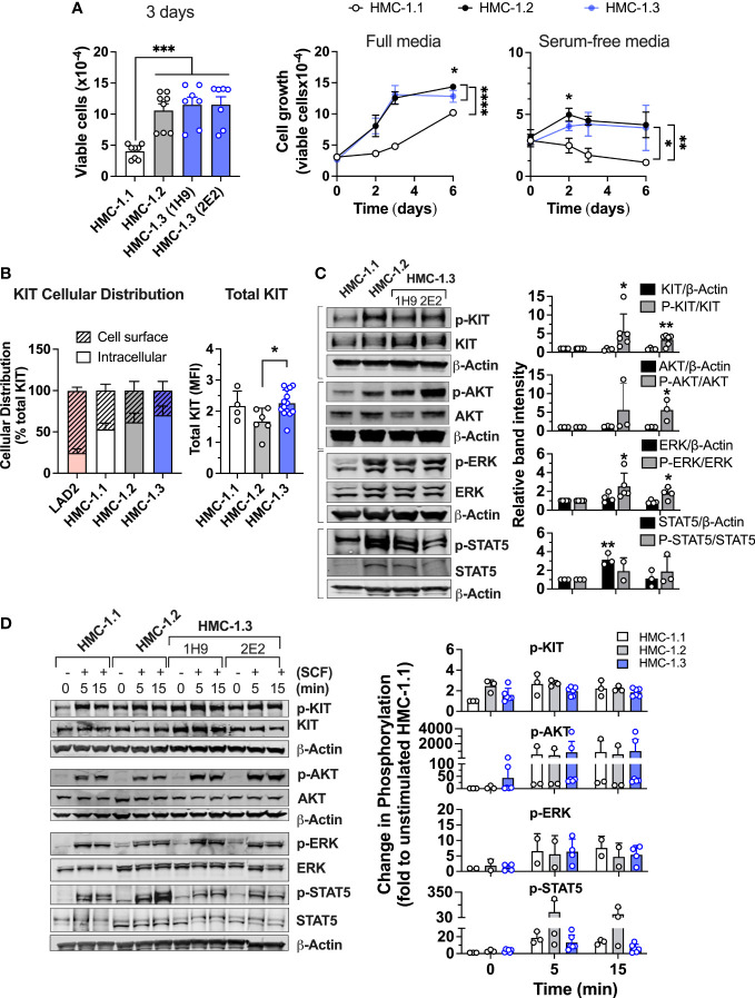
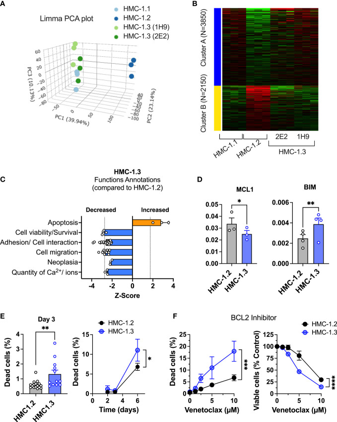
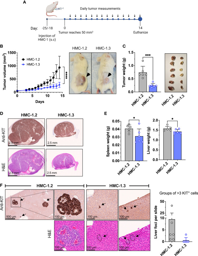
The HMC-1.2 human mast cell (huMC) line is often employed in the study of attributes of neoplastic huMCs as found in patients with mastocytosis and their sensitivity to interventional drugs and . HMC-1.2 cells express constitutively active KIT, an essential growth factor receptor for huMC survival and function, due to the presence of two oncogenic mutations (D816V and V560G). However, systemic mastocytosis is commonly associated with a single D816V-KIT mutation. The functional consequences of the coexisting KIT mutations in HMC-1.2 cells are unknown. We used CRISPR/Cas9-engineering to reverse the V560G mutation in HMC-1.2 cells, resulting in a subline (HMC-1.3) with a single mono-allelic D816V-KIT variant. Transcriptome analyses predicted reduced activity in pathways involved in survival, cell-to-cell adhesion, and neoplasia in HMC-1.3 compared to HMC-1.2 cells, with differences in expression of molecular components and cell surface markers. Consistently, subcutaneous inoculation of HMC-1.3 into mice produced significantly smaller tumors than HMC-1.2 cells, and in colony assays, HMC-1.3 formed less numerous and smaller colonies than HMC-1.2 cells. However, in liquid culture conditions, the growth of HMC-1.2 and HMC-1.3 cells was comparable. Phosphorylation levels of ERK1/2, AKT and STAT5, representing pathways associated with constitutive oncogenic KIT signaling, were also similar between HMC-1.2 and HMC-1.3 cells. Despite these similarities in liquid culture, survival of HMC-1.3 cells was diminished in response to various pharmacological inhibitors, including tyrosine kinase inhibitors used clinically for treatment of advanced systemic mastocytosis, and JAK2 and BCL2 inhibitors, making HMC-1.3 more susceptible to these drugs than HMC-1.2 cells. Our study thus reveals that the additional V560G-KIT oncogenic variant in HMC-1.2 cells modifies transcriptional programs induced by D816V-KIT, confers a survival advantage, alters sensitivity to interventional drugs, and increases the tumorigenicity, suggesting that engineered huMCs with a single D816V-KIT variant may represent an improved preclinical model for mastocytosis.
HMC-1.2 人肥大细胞(huMC)系常用于研究肥大细胞肿瘤患者中发现的肿瘤性 huMC 的特性及其对干预性药物的敏感性。由于存在两种致癌突变(D816V 和 V560G),HMC-1.2 细胞持续表达构成性激活的 KIT,这是 huMC 存活和功能的必需生长因子受体。然而,系统性肥大细胞增多症通常与单个 D816V-KIT 突变相关。HMC-1.2 细胞中共同存在的 KIT 突变的功能后果尚不清楚。我们使用 CRISPR/Cas9 工程技术逆转 HMC-1.2 细胞中的 V560G 突变,产生了一个亚系(HMC-1.3),其中存在单个单等位基因 D816V-KIT 变体。转录组分析预测,与 HMC-1.2 细胞相比,HMC-1.3 中涉及存活、细胞间粘附和肿瘤发生的途径活性降低,分子成分和细胞表面标志物的表达存在差异。一致地,HMC-1.3 细胞的皮下接种在小鼠中产生的肿瘤明显小于 HMC-1.2 细胞,并且在集落测定中,HMC-1.3 细胞形成的集落数量较少且较小。然而,在液体培养条件下,HMC-1.2 和 HMC-1.3 细胞的生长相似。ERK1/2、AKT 和 STAT5 的磷酸化水平,代表与组成性致癌 KIT 信号相关的途径,在 HMC-1.2 和 HMC-1.3 细胞之间也相似。尽管在液体培养中有这些相似之处,但 HMC-1.3 细胞对各种药理抑制剂的存活能力降低,包括临床上用于治疗晚期系统性肥大细胞增多症的酪氨酸激酶抑制剂以及 JAK2 和 BCL2 抑制剂,使 HMC-1.3 细胞对这些药物比 HMC-1.2 细胞更敏感。因此,我们的研究表明,HMC-1.2 细胞中额外的 V560G-KIT 致癌变体改变了由 D816V-KIT 诱导的转录程序,赋予了生存优势,改变了对干预性药物的敏感性,并增加了致瘤性,表明具有单个 D816V-KIT 变体的工程化 huMC 可能代表一种改进的肥大细胞增多症的临床前模型。