Division of Gastroenterology and Hepatology, Department of Internal Medicine, Faculty of Medicine, University of Miyazaki, Japan.
Department of Pathology, Faculty of Medicine, University of Miyazaki, Japan.
Intern Med. 2023 Nov 1;62(21):3143-3149. doi: 10.2169/internalmedicine.0967-22. Epub 2023 Apr 7.
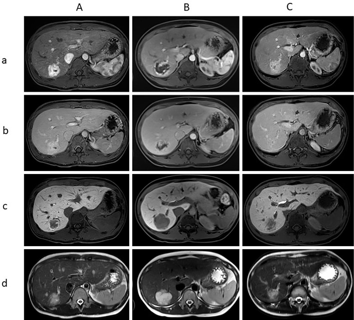
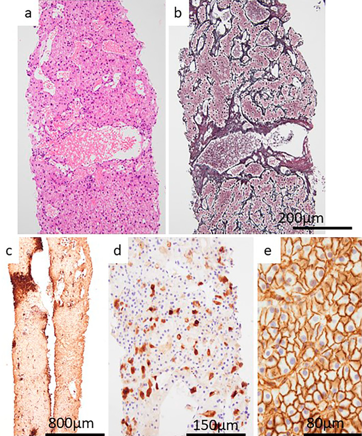
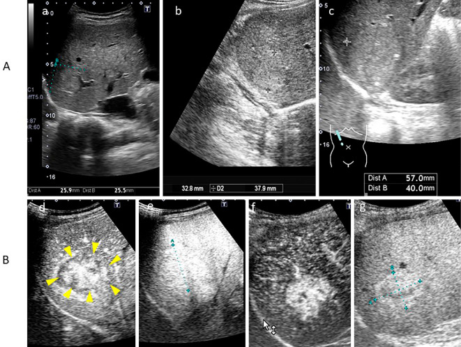
We reported a notable case of inflammatory hepatocellular adenoma that grew during pregnancy, consequently changing its appearance on magnetic resonance imaging remarkably. A 5-months-pregnant 35-year-old woman presented with a 37-mm liver nodule that had been diagnosed as focal nodular hyperplasia 3 years earlier. She had never used oral contraceptives. After 2 months, the nodule grew to 57 mm. The patient delivered a full-term infant without complications. Gadolinium-ethoxybenzyl-diethylenetriamine pentaacetic acid-enhanced magnetic resonance imaging performed after delivery revealed markedly different findings compared with the first images. A liver biopsy was performed, and the tumor was diagnosed as inflammatory hepatocellular adenoma.
我们报告了一例妊娠期间生长的炎症性肝细胞腺瘤的显著病例,其磁共振成像表现显著改变。一位 35 岁的 5 个月孕妇,3 年前曾被诊断为局灶性结节性增生,出现一个 37mm 的肝脏结节。她从未服用过口服避孕药。2 个月后,结节长到 57mm。患者顺利分娩,无并发症。分娩后行钆喷替酸葡甲胺增强磁共振成像,与首次图像相比,发现明显不同的结果。行肝活检,诊断为炎症性肝细胞腺瘤。