Wang Meixia, Cheng Xiaoyu, Shi Qianru, Xu Bo, Hou Xiaoxia, Zhao Huimin, Gui Qian, Wu Guanhui, Dong Xiaofeng, Xu Qinrong, Shen Mingqiang, Cheng Qingzhang, Xue Shouru, Feng Hongxuan, Ding Zhiliang
Department of Neurology, Suzhou Hospital Affiliated to Nanjing Medical University, Suzhou Municipal Hospital, Suzhou, China.
Department of Neurology and Clinical Research Center of Neurological Disease, The Second Affiliated Hospital of Soochow University, Suzhou, China.
Front Hum Neurosci. 2023 Mar 22;17:1142408. doi: 10.3389/fnhum.2023.1142408. eCollection 2023.
Accumulating evidence shows that epilepsy is a disease caused by brain network dysfunction. This study explored changes in brain network structure in epilepsy patients based on graph analysis of diffusion tensor imaging data.
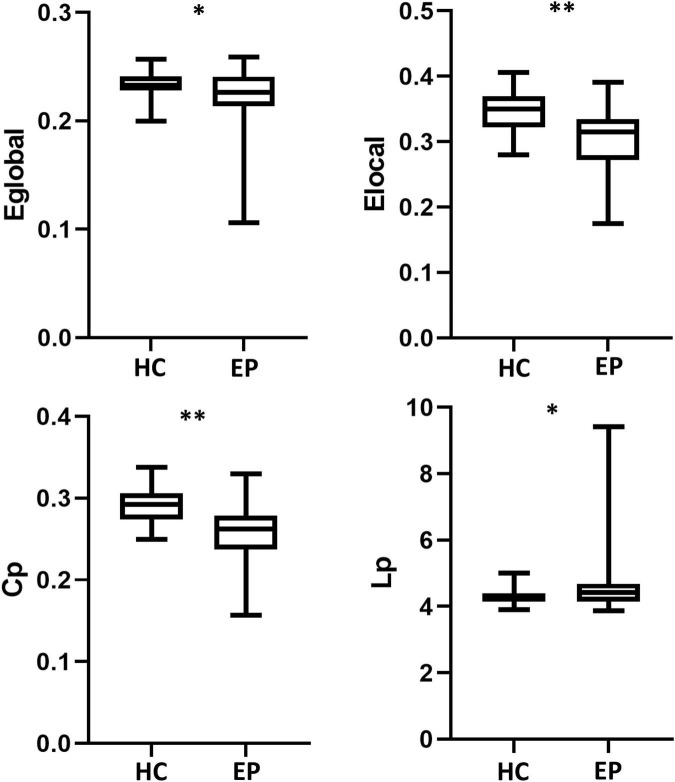
The brain structure networks of 42 healthy control individuals and 26 epilepsy patients were constructed. Using graph theory analysis, global and local network topology parameters of the brain structure network were calculated, and changes in global and local characteristics of the brain network in epilepsy patients were quantitatively analyzed.
Compared with the healthy control group, the epilepsy patient group showed lower global efficiency, local efficiency, clustering coefficient, and a longer shortest path length. Both healthy control individuals and epilepsy patients showed small-world attributes, with no significant difference between groups. The epilepsy patient group showed lower nodal local efficiency and nodal clustering coefficient in the right olfactory cortex and right rectus and lower nodal degree centrality in the right olfactory cortex and the left paracentral lobular compared with the healthy control group. In addition, the epilepsy patient group showed a smaller fiber number of edges in specific regions of the frontal lobe, temporal lobe, and default mode network, indicating reduced connection strength.
Epilepsy patients exhibited lower global and local brain network properties as well as reduced white matter fiber connectivity in key brain regions. These findings further support the idea that epilepsy is a brain network disorder.
越来越多的证据表明,癫痫是一种由脑网络功能障碍引起的疾病。本研究基于扩散张量成像数据的图谱分析,探讨癫痫患者脑网络结构的变化。
构建42名健康对照个体和26名癫痫患者的脑结构网络。采用图谱理论分析方法,计算脑结构网络的全局和局部网络拓扑参数,定量分析癫痫患者脑网络全局和局部特征的变化。
与健康对照组相比,癫痫患者组的全局效率、局部效率、聚类系数较低,最短路径长度较长。健康对照个体和癫痫患者均表现出小世界属性,两组之间无显著差异。与健康对照组相比,癫痫患者组右侧嗅皮质和右侧直肌的节点局部效率和节点聚类系数较低,右侧嗅皮质和左侧中央旁小叶的节点度中心性较低。此外,癫痫患者组额叶、颞叶和默认模式网络特定区域的纤维边数较少,表明连接强度降低。
癫痫患者表现出较低的全局和局部脑网络特性,以及关键脑区白质纤维连接性降低。这些发现进一步支持了癫痫是一种脑网络疾病的观点。