Zhu Hongyu, Chen Peng, Dong Guozhang, Meng Fanchen, Xia Zhijun, You Jing, Kong Xiangru, Wu Jintao, Yuan Fangwei, Yu Xinyu, Sun Qinhong, Ji Jinfu, Wang Siwei, Liu Tongyan, Xu Lin
Department of Thoracic Surgery, the Affiliated Cancer Hospital of Nanjing Medical University, Jiangsu Cancer Hospital, Jiangsu Institute of Cancer Research, Jiangsu Key Laboratory of Molecular and Translation Cancer Research, Nanjing 210009, China.
Zhongguo Fei Ai Za Zhi. 2023 Mar 20;26(3):204-216. doi: 10.3779/j.issn.1009-3419.2023.101.07.
The morbidity and mortality rates of lung cancer remain high worldwide, and lung adenocarcinoma is one of the most important tissue subtypes of lung cancer. Epidermal growth factor receptor (EGFR) mutation is an important driver gene mutation for lung adenocarcinoma. In recent years, immune checkpoint inhibitors (ICIs), such as programmed cell death 1 (PD-1) and programmed cell death ligand 1 (PD-L1) inhibitors, have achieved remarkable efficacy in some lung cancer patients. Patients with EGFR mutations enjoyed limited benefits from immunotherapy according to recent studies. This study aimed to explore the relationship between EGFR mutation status and the spatial distribution as well as infiltration number of various immune cells in patients with EGFR mutant lung adenocarcinoma.
This study included 62 lung adenocarcinoma patients who underwent surgery. Through multi-point sampling of surgically removed tumor tissues in different areas, 223 tumor tissue samples were finally obtained. We aquired EGFR mutations status including variant allele frequency (VAF) and mutation subtype in each tumor tissue by genetic test. Afterwards, hematoxylin-eosin (HE) staining, immunohistochemical staining and multiplex fluorescence immunohistochemistry staining have been performed, therefore the infiltration of various immune cells and the distribution of tertiary lymphoid structure (TLS) in tumor tissues were obtained by calculating the immunohistochemical score.
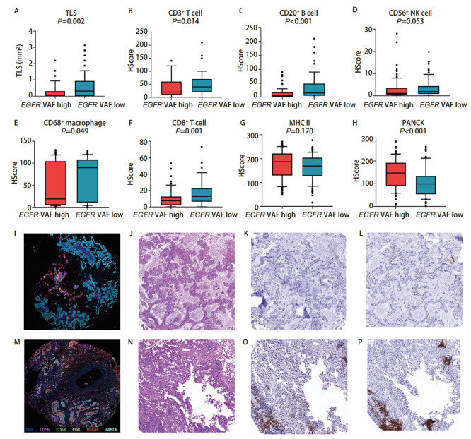
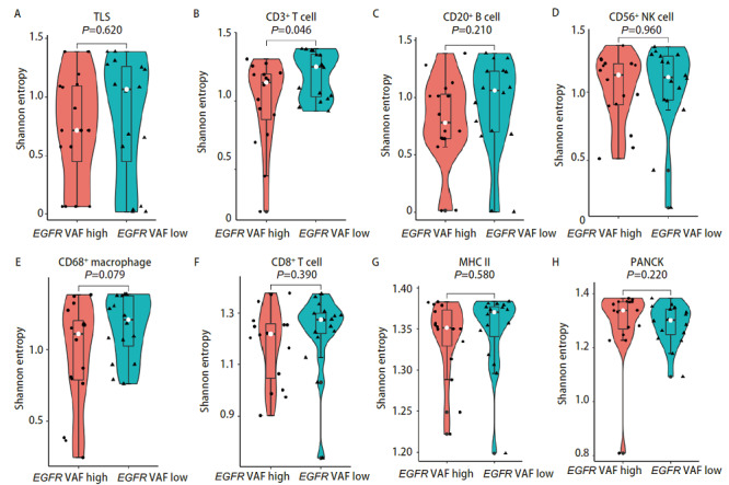
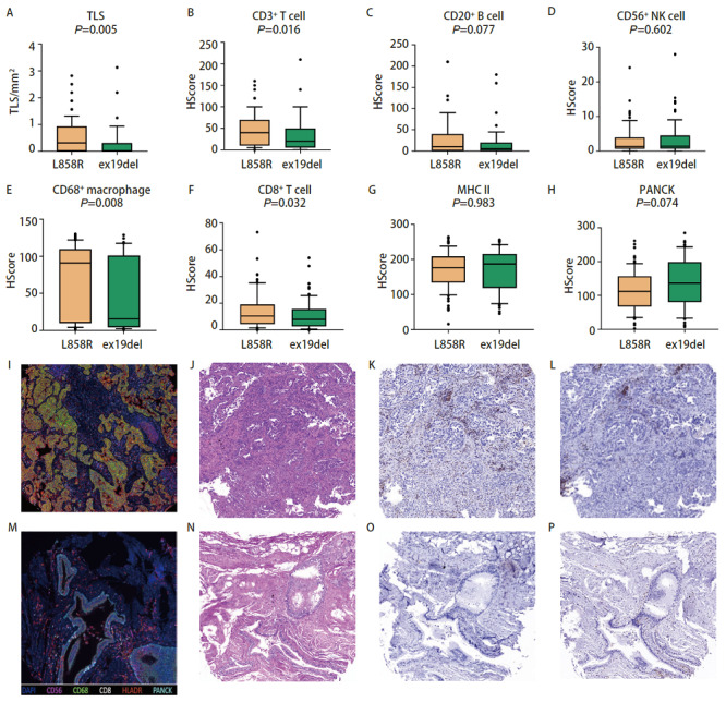
Compared with EGFR wild-type patients, patients with EGFR-mutant lung adenocarcinoma had more infiltration of CD68+ macrophages and major histocompatibility complex (MHC) class II antigen-presenting cells and higher spatial distribution heterogeneity of MHC class II antigen presenting cells in tumor tissues, while CD56+ natural killer cells and CD8+ T cells had lower infiltration. Tumor tissues with higher EGFR VAF were associated with lower cell infiltration such as CD3+ T cells, CD20+ B cells, CD56+ natural killer cells, CD68+ macrophages, CD8+ T cells, and only CD3+ T cells showed a lower spatial distribution heterogeneity. For the two common subtypes of EGFR mutations in Chinese population, tumor tissues with EGFR exon 19 deletion mutations have lower immune cell infiltration but higher spatial distribution heterogeneity of CD3+ T cells, CD56+ natural killer cells, CD68+ macrophages, and CD8+ T cells than that in EGFR exon 21 L858R mutant tumor tissues. Prognostic analysis found that patients with EGFR mutations with high degree of CD3+ T cells, CD20+ B cell infiltration and larger numbers of TLS formation and high spatial distribution heterogeneity of CD8+ T cell had longer disease-free survival.
EGFR-mutated lung adenocarcinoma had a unique "non-inflammatory" tumor microenvironment with low infiltration of immune cells, and there was also heterogeneity in the tumor microenvironment among the tumors with different mutation subtypes and mutation abundance. These differences were not only reflected in the number but also the spatial distribution of immune cell infiltration. Hence, further studies on the immune microenvironment of EGFR-mutant lung adenocarcinoma were of great significance for improving the efficacy of immunotherapy in EGFR-mutant lung adenocarcinoma patients in the future.
肺癌的发病率和死亡率在全球范围内仍然很高,肺腺癌是肺癌最重要的组织学亚型之一。表皮生长因子受体(EGFR)突变是肺腺癌的重要驱动基因突变。近年来,免疫检查点抑制剂(ICI),如程序性细胞死亡蛋白1(PD-1)和程序性细胞死亡配体1(PD-L1)抑制剂,在一些肺癌患者中取得了显著疗效。根据最近的研究,EGFR突变患者从免疫治疗中获益有限。本研究旨在探讨EGFR突变状态与EGFR突变型肺腺癌患者各种免疫细胞的空间分布及浸润数量之间的关系。
本研究纳入62例接受手术的肺腺癌患者。通过对手术切除的肿瘤组织进行不同区域的多点采样,最终获得223个肿瘤组织样本。我们通过基因检测获得每个肿瘤组织的EGFR突变状态,包括变异等位基因频率(VAF)和突变亚型。之后,进行苏木精-伊红(HE)染色、免疫组织化学染色和多重荧光免疫组织化学染色,通过计算免疫组织化学评分获得肿瘤组织中各种免疫细胞的浸润情况和三级淋巴结构(TLS)的分布。
与EGFR野生型患者相比,EGFR突变型肺腺癌患者的肿瘤组织中CD68+巨噬细胞和主要组织相容性复合体(MHC)II类抗原呈递细胞浸润更多,MHC II类抗原呈递细胞的空间分布异质性更高,而CD56+自然杀伤细胞和CD8+T细胞浸润较低。EGFR VAF较高的肿瘤组织与较低的细胞浸润相关,如CD3+T细胞、CD20+B细胞、CD56+自然杀伤细胞、CD68+巨噬细胞、CD8+T细胞,只有CD3+T细胞表现出较低的空间分布异质性。对于中国人群中两种常见的EGFR突变亚型,与EGFR外显子21 L858R突变肿瘤组织相比,EGFR外显子19缺失突变的肿瘤组织免疫细胞浸润较低,但CD3+T细胞、CD56+自然杀伤细胞、CD68+巨噬细胞和CD8+T细胞的空间分布异质性较高。预后分析发现,CD3+T细胞、CD20+B细胞浸润程度高、TLS形成数量多且CD8+T细胞空间分布异质性高的EGFR突变患者无病生存期更长。
EGFR突变型肺腺癌具有独特的“非炎症性”肿瘤微环境,免疫细胞浸润低,不同突变亚型和突变丰度的肿瘤之间肿瘤微环境也存在异质性。这些差异不仅体现在免疫细胞浸润的数量上,还体现在空间分布上。因此,进一步研究EGFR突变型肺腺癌的免疫微环境对于未来提高EGFR突变型肺腺癌患者的免疫治疗疗效具有重要意义。