Yong Loo Lin School of Medicine, National University of Singapore, Singapore, 169856, Singapore.
Department of Anatomical Pathology, Singapore General Hospital, Singapore, 169856, Singapore.
Cancer Commun (Lond). 2020 Apr;40(4):135-153. doi: 10.1002/cac2.12023. Epub 2020 Apr 17.
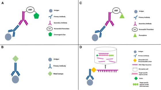
Conventional immunohistochemistry (IHC) is a widely used diagnostic technique in tissue pathology. However, this technique is associated with a number of limitations, including high inter-observer variability and the capacity to label only one marker per tissue section. This review details various highly multiplexed techniques that have emerged to circumvent these constraints, allowing simultaneous detection of multiple markers on a single tissue section and the comprehensive study of cell composition, cellular functional and cell-cell interactions. Among these techniques, multiplex Immunohistochemistry/Immunofluorescence (mIHC/IF) has emerged to be particularly promising. mIHC/IF provides high-throughput multiplex staining and standardized quantitative analysis for highly reproducible, efficient and cost-effective tissue studies. This technique has immediate potential for translational research and clinical practice, particularly in the era of cancer immunotherapy.
传统的免疫组织化学(IHC)是组织病理学中广泛使用的诊断技术。然而,该技术存在许多局限性,包括观察者间变异性高,以及每次组织切片只能标记一个标记物。本综述详细介绍了各种新兴的高度多重化技术,这些技术可克服这些限制,允许在单个组织切片上同时检测多个标记物,并全面研究细胞组成、细胞功能和细胞-细胞相互作用。在这些技术中,多重免疫组织化学/免疫荧光(mIHC/IF)技术尤其有前途。mIHC/IF 提供高通量多重染色和标准化定量分析,具有高度可重复性、高效和经济有效的组织研究。该技术在转化研究和临床实践中具有直接的应用潜力,特别是在癌症免疫治疗时代。