Foesleitner Olivia, Knop Karl Christian, Lindenau Matthias, Preisner Fabian, Bäumer Philipp, Heiland Sabine, Bendszus Martin, Kronlage Moritz
Department of Neuroradiology, Heidelberg University Hospital, Im Neuenheimer Feld 400, 69120 Heidelberg, Germany.
Neurologie Neuer Wall Dr. Bredow & Partner, Neuer Wall 19, 20354 Hamburg, Germany.
Diagnostics (Basel). 2023 Mar 25;13(7):1237. doi: 10.3390/diagnostics13071237.
The aim of this study was to assess the phenotype of multifocal motor neuropathy (MMN) and amyotrophic lateral sclerosis (ALS) in quantitative MR neurography.
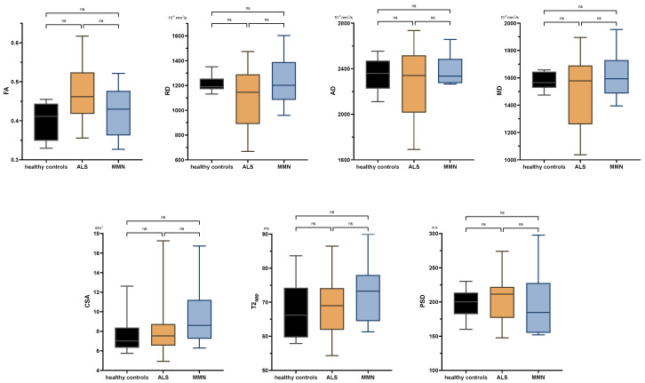
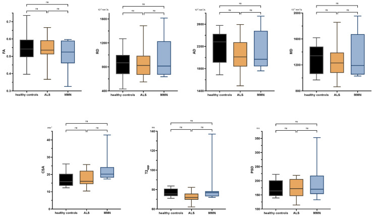
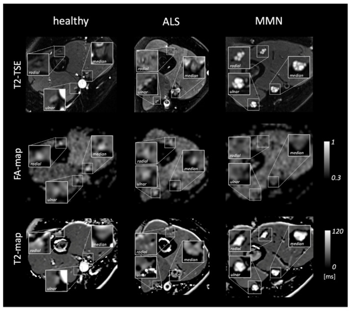
In this prospective study, 22 patients with ALS, 8 patients with MMN, and 10 healthy volunteers were examined with 3T MR neurography, using a high-resolution fat-saturated T2-weighted sequence, diffusion-tensor imaging (DTI), and a multi-echo T2-relaxometry sequence. The quantitative biomarkers fractional anisotropy (FA), radial and axial diffusivity (RD, AD), mean diffusivity (MD), cross-sectional area (CSA), T2-relaxation time, and proton spin density (PSD) were measured in the tibial nerve at the thigh and calf, and in the median, radial, and ulnar nerves at the mid-upper arm.
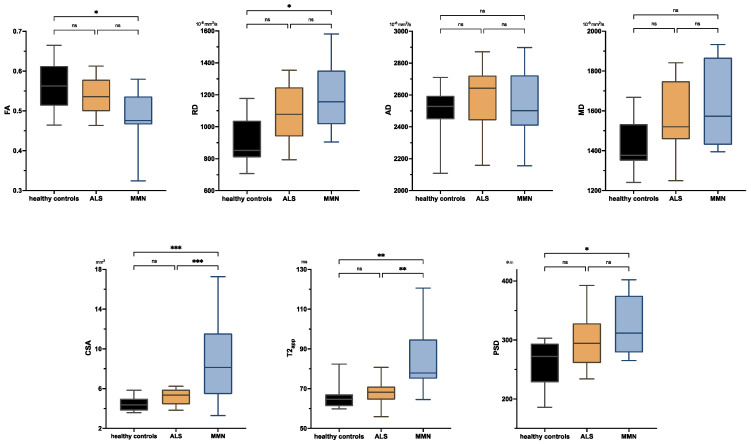
MMN showed a characteristic imaging pattern of decreased FA ( = 0.018), increased RD ( = 0.014), increased CSA ( < 0.001), increased T2-relaxation time ( < 0.001), and increased PSD ( = 0.025) in the upper arm nerves compared to ALS and controls. ALS patients did not differ from controls in any imaging marker, nor were there any group differences in the tibial nerve ( > 0.05).
MMN shows a characteristic pattern of quantitative DTI and T2-relaxometry parameters in the upper-arm nerves, primarily indicating demyelination. Peripheral nerve changes in ALS seem to be below the detection level of current state-of-the-art quantitative MR neurography.
本研究旨在通过定量磁共振神经成像评估多灶性运动神经病(MMN)和肌萎缩侧索硬化症(ALS)的表型。
在这项前瞻性研究中,对22例ALS患者、8例MMN患者和10名健康志愿者进行了3T磁共振神经成像检查,使用高分辨率脂肪饱和T2加权序列、扩散张量成像(DTI)和多回波T2弛豫测量序列。在大腿和小腿的胫神经以及上臂中部的正中神经、桡神经和尺神经中测量定量生物标志物各向异性分数(FA)、径向和轴向扩散率(RD、AD)、平均扩散率(MD)、横截面积(CSA)、T2弛豫时间和质子自旋密度(PSD)。
与ALS患者和对照组相比,MMN在上臂神经中表现出特征性的成像模式,即FA降低( = 0.018)、RD增加( = 0.014)、CSA增加( < 0.001)、T2弛豫时间增加( < 0.001)和PSD增加( = 0.025)。ALS患者在任何成像标志物方面与对照组均无差异,胫神经也无组间差异( > 0.05)。
MMN在上臂神经中表现出定量DTI和T2弛豫测量参数的特征性模式,主要提示脱髓鞘。ALS的周围神经变化似乎低于当前先进的定量磁共振神经成像的检测水平。