Kudsieh Bachar, Fernández-Vigo José Ignacio, Flores-Moreno Ignacio, Ruiz-Medrano Jorge, Garcia-Zamora Maria, Samaan Muhsen, Ruiz-Moreno Jose Maria
Department of Ophthalmology, University Hospital Puerta De Hierro Majadahonda, 28220 Madrid, Spain.
Centro Internacional de Oftalmologia Avanzada, 28010 Madrid, Spain.
J Clin Med. 2023 Mar 29;12(7):2592. doi: 10.3390/jcm12072592.
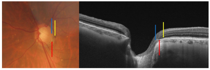
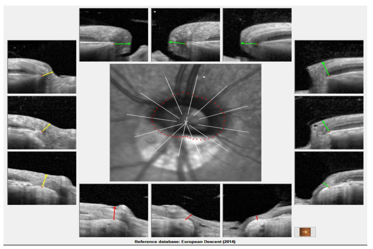
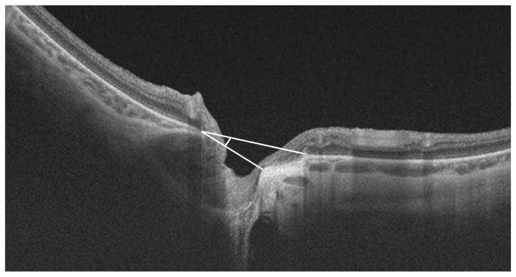
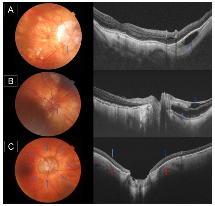
Glaucoma diagnosis in highly myopic subjects by optic nerve head (ONH) imaging is challenging as it is difficult to distinguish structural defects related to glaucoma from myopia-related defects in these subjects. Optical coherence tomography (OCT) has evolved to become a routine examination at present, providing key information in the assessment of glaucoma based on the study of the ONH. However, the correct segmentation and interpretation of the ONH data employing OCT is still a challenge in highly myopic patients. High-resolution OCT images can help qualitatively and quantitatively describe the structural characteristics and anatomical changes in highly myopic subjects with and without glaucoma. The ONH and peripapillary area can be analyzed to measure the myopic atrophic-related zone, the existence of intrachoroidal cavitation, staphyloma, and ONH pits by OCT. Similarly, the lamina cribosa observed in the OCT images may reveal anatomical changes that justify visual defects. Several quantitative parameters of the ONH obtained from OCT images were proposed to predict the progression of visual defects in glaucoma subjects. Additionally, OCT images help identify factors that may negatively influence the measurement of the retinal nerve fiber layer (RNFL) and provide better analysis using new parameters, such as Bruch's Membrane Opening-Minimum Rim Width, which serves as an alternative to RNFL measurements in highly myopic subjects due to its superior diagnostic ability.
通过视神经乳头(ONH)成像对高度近视患者进行青光眼诊断具有挑战性,因为在这些患者中,很难将与青光眼相关的结构缺陷与近视相关的缺陷区分开来。光学相干断层扫描(OCT)目前已发展成为一项常规检查,通过对视神经乳头的研究,为青光眼评估提供关键信息。然而,在高度近视患者中,使用OCT对视神经乳头数据进行正确的分割和解读仍然是一项挑战。高分辨率OCT图像有助于定性和定量描述患有和未患有青光眼的高度近视患者的结构特征和解剖学变化。可以通过OCT分析视神经乳头和视乳头周围区域,以测量近视萎缩相关区域、脉络膜内空洞、葡萄肿和视神经乳头凹陷的存在情况。同样,在OCT图像中观察到的筛板可能揭示出导致视觉缺陷的解剖学变化。有人提出了从OCT图像中获得的视神经乳头的几个定量参数,以预测青光眼患者视觉缺陷的进展。此外,OCT图像有助于识别可能对视神经纤维层(RNFL)测量产生负面影响的因素,并使用新的参数(如Bruch膜开口 - 最小边缘宽度)进行更好的分析,由于其卓越的诊断能力,该参数可作为高度近视患者RNFL测量的替代方法。