• 文献检索
  • 文档翻译
  • 深度研究
  • 学术资讯
  • Suppr Zotero 插件Zotero 插件
  • 邀请有礼
  • 套餐&价格
  • 历史记录
应用&插件
Suppr Zotero 插件Zotero 插件浏览器插件Mac 客户端Windows 客户端微信小程序
定价
高级版会员购买积分包购买API积分包
服务
文献检索文档翻译深度研究API 文档MCP 服务
关于我们
关于 Suppr公司介绍联系我们用户协议隐私条款
关注我们

Suppr 超能文献

核心技术专利:CN118964589B侵权必究
粤ICP备2023148730 号-1Suppr @ 2026

文献检索

告别复杂PubMed语法,用中文像聊天一样搜索,搜遍4000万医学文献。AI智能推荐,让科研检索更轻松。

立即免费搜索

文件翻译

保留排版,准确专业,支持PDF/Word/PPT等文件格式,支持 12+语言互译。

免费翻译文档

深度研究

AI帮你快速写综述,25分钟生成高质量综述,智能提取关键信息,辅助科研写作。

立即免费体验

猪眼表面上皮微环境的特征。

Characterization of Porcine Ocular Surface Epithelial Microenvironment.

机构信息

Eye Center, Medical Center, Faculty of Medicine, University of Freiburg, Killianstrasse 5, 79106 Freiburg, Germany.

CEMT-Freiburg, Experimental Surgery, Hospital-Medical Center, Faculty of Medicine, University of Freiburg, Breisacher Str. 66, 79106 Freiburg, Germany.

出版信息

Int J Mol Sci. 2023 Apr 19;24(8):7543. doi: 10.3390/ijms24087543.

DOI:10.3390/ijms24087543
PMID:37108705
原文链接:https://pmc.ncbi.nlm.nih.gov/articles/PMC10145510/
Abstract

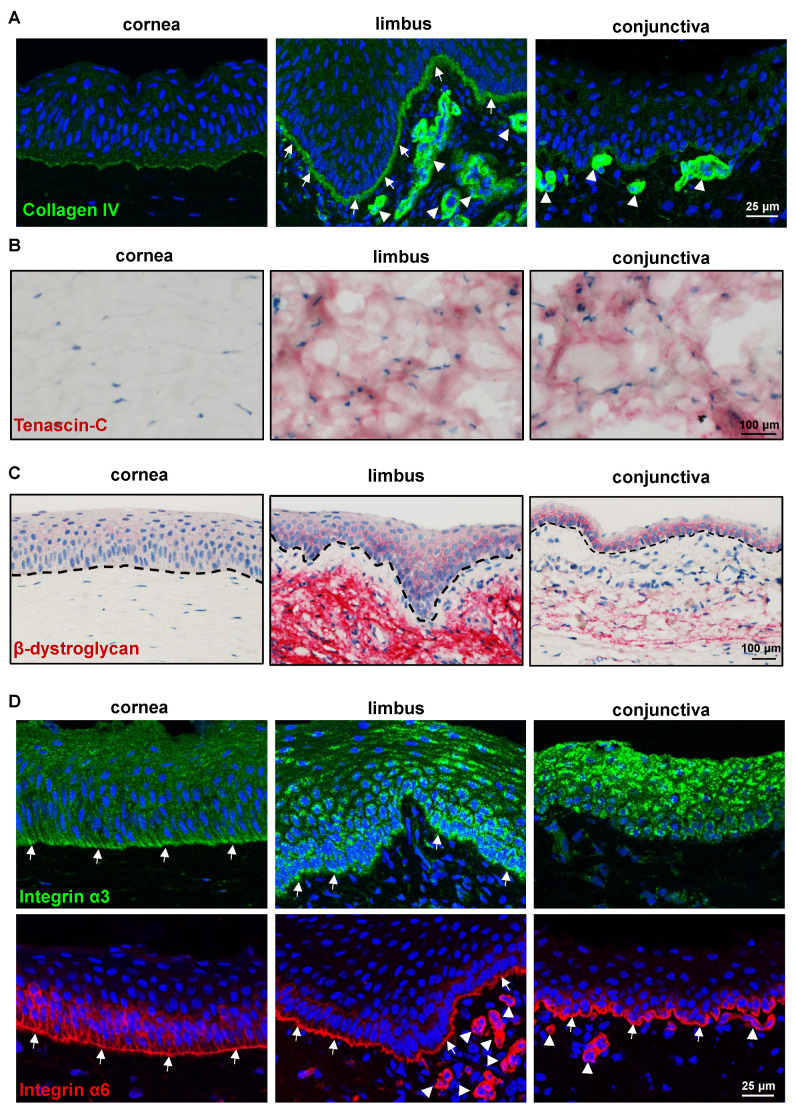
The porcine ocular surface is used as a model of the human ocular surface; however, a detailed characterization of the porcine ocular surface has not been documented. This is due, in part, to the scarcity of antibodies produced specifically against the porcine ocular surface cell types or structures. We performed a histological and immunohistochemical investigation on frozen and formalin-fixed, paraffin-embedded ocular surface tissue from domestic pigs using a panel of 41 different antibodies related to epithelial progenitor/differentiation phenotypes, extracellular matrix and associated molecules, and various niche cell types. Our observations suggested that the Bowman's layer is not evident in the cornea; the deep invaginations of the limbal epithelium in the limbal zone are analogous to the limbal interpalisade crypts of human limbal tissue; and the presence of goblet cells in the bulbar conjunctiva. Immunohistochemistry analysis revealed that the epithelial progenitor markers cytokeratin (CK)15, CK14, p63α, and P-cadherin were expressed in both the limbal and conjunctival basal epithelium, whereas the basal cells of the limbal and conjunctival epithelium did not stain for CK3, CK12, E-cadherin, and CK13. Antibodies detecting marker proteins related to the extracellular matrix (collagen IV, Tenascin-C), cell-matrix adhesion (β-dystroglycan, integrin α3 and α6), mesenchymal cells (vimentin, CD90, CD44), neurons (neurofilament), immune cells (HLA-ABC; HLA-DR, CD1, CD4, CD14), vasculature (von Willebrand factor), and melanocytes (SRY-homeobox-10, human melanoma black-45, Tyrosinase) on the normal human ocular surface demonstrated similar immunoreactivity on the normal porcine ocular surface. Only a few antibodies (directed against N-cadherin, fibronectin, agrin, laminin α3 and α5, melan-A) appeared unreactive on porcine tissues. Our findings characterize the main immunohistochemical properties of the porcine ocular surface and provide a morphological and immunohistochemical basis useful to research using porcine models. Furthermore, the analyzed porcine ocular structures are similar to those of humans, confirming the potential usefulness of pig eyes to study ocular surface physiology and pathophysiology.

摘要

猪的眼表面被用作人眼表面的模型;然而,尚未详细描述猪眼表面。这部分是由于缺乏专门针对猪眼表面细胞类型或结构的抗体。我们使用一组 41 种不同的抗体,对来自家猪的冷冻和福尔马林固定、石蜡包埋的眼表面组织进行了组织学和免疫组织化学研究,这些抗体与上皮祖细胞/分化表型、细胞外基质和相关分子以及各种生态位细胞类型有关。我们的观察结果表明,Bowman 层在角膜中不明显;角膜缘区的缘上皮的深层内陷类似于人角膜缘组织的缘间栅栏隐窝;以及在球结膜中存在杯状细胞。免疫组织化学分析显示,上皮祖细胞标志物角蛋白 (CK)15、CK14、p63α 和 P-钙黏蛋白在角膜缘和结膜基底上皮中均有表达,而角膜缘和结膜上皮的基底细胞则不染色 CK3、CK12、E-钙黏蛋白和 CK13。检测与细胞外基质 (胶原 IV、Tenascin-C)、细胞-基质黏附 (β- 肌营养不良蛋白、整合素 α3 和 α6)、间充质细胞 (波形蛋白、CD90、CD44)、神经元 (神经丝)、免疫细胞 (HLA-ABC;HLA-DR、CD1、CD4、CD14)、血管 (血管性血友病因子) 和黑素细胞 (SRY-同源盒-10、人黑素瘤黑 45、酪氨酸酶) 的相关标记蛋白的抗体在正常人类眼表面表现出类似的免疫反应性,也存在于正常猪眼表面。只有少数几种抗体(针对 N-钙黏蛋白、纤连蛋白、聚集蛋白、层粘连蛋白 α3 和 α5、黑素-A)在猪组织中似乎没有反应。我们的发现描述了猪眼表面的主要免疫组织化学特性,并为使用猪模型进行研究提供了形态学和免疫组织化学基础。此外,分析的猪眼结构与人类相似,证实了猪眼在研究眼表面生理学和病理生理学方面的潜在有用性。

https://cdn.ncbi.nlm.nih.gov/pmc/blobs/7c3e/10145510/1cbce6e2a2e5/ijms-24-07543-g008.jpg
https://cdn.ncbi.nlm.nih.gov/pmc/blobs/7c3e/10145510/25d89b0134b3/ijms-24-07543-g001.jpg
https://cdn.ncbi.nlm.nih.gov/pmc/blobs/7c3e/10145510/40b8f35d90a1/ijms-24-07543-g002.jpg
https://cdn.ncbi.nlm.nih.gov/pmc/blobs/7c3e/10145510/46f2229dba95/ijms-24-07543-g003.jpg
https://cdn.ncbi.nlm.nih.gov/pmc/blobs/7c3e/10145510/5b56352064e9/ijms-24-07543-g004.jpg
https://cdn.ncbi.nlm.nih.gov/pmc/blobs/7c3e/10145510/4a799ece4070/ijms-24-07543-g005.jpg
https://cdn.ncbi.nlm.nih.gov/pmc/blobs/7c3e/10145510/f9428fe54eab/ijms-24-07543-g006.jpg
https://cdn.ncbi.nlm.nih.gov/pmc/blobs/7c3e/10145510/be5f39472aa3/ijms-24-07543-g007.jpg
https://cdn.ncbi.nlm.nih.gov/pmc/blobs/7c3e/10145510/1cbce6e2a2e5/ijms-24-07543-g008.jpg

相似文献

1
Characterization of Porcine Ocular Surface Epithelial Microenvironment.猪眼表面上皮微环境的特征。
Int J Mol Sci. 2023 Apr 19;24(8):7543. doi: 10.3390/ijms24087543.
2
Characterization of extracellular matrix components in the limbal epithelial stem cell compartment.角膜缘上皮干细胞区室中细胞外基质成分的特征分析。
Exp Eye Res. 2007 Dec;85(6):845-60. doi: 10.1016/j.exer.2007.08.020. Epub 2007 Sep 2.
3
A single cell atlas of human cornea that defines its development, limbal progenitor cells and their interactions with the immune cells.人类眼角膜单细胞图谱,定义其发育、角膜缘祖细胞及其与免疫细胞的相互作用。
Ocul Surf. 2021 Jul;21:279-298. doi: 10.1016/j.jtos.2021.03.010. Epub 2021 Apr 16.
4
The porcine limbal epithelial stem cell niche as a new model for the study of transplanted tissue-engineered human limbal epithelial cells.猪角膜缘上皮干细胞龛作为研究移植组织工程化人角膜缘上皮细胞的新模型。
Tissue Eng Part A. 2011 Mar;17(5-6):741-50. doi: 10.1089/ten.TEA.2010.0343. Epub 2011 Jan 16.
5
Histological and immunohistochemical characterization of the porcine ocular surface.猪眼表面的组织学和免疫组织化学特征。
PLoS One. 2020 Jan 13;15(1):e0227732. doi: 10.1371/journal.pone.0227732. eCollection 2020.
6
Cytokeratin 8 is expressed in human corneoconjunctival epithelium, particularly in limbal epithelial cells.细胞角蛋白 8 表达于人类的角膜结膜上皮,尤其是在角膜缘上皮细胞中。
Invest Ophthalmol Vis Sci. 2011 Feb 9;52(2):787-94. doi: 10.1167/iovs.10-5489.
7
Influence of Organ Culture on the Characteristics of the Human Limbal Stem Cell Niche.器官培养对人角膜缘干细胞龛特征的影响。
Int J Mol Sci. 2023 Nov 28;24(23):16856. doi: 10.3390/ijms242316856.
8
Differential Distribution of Laminin N-Terminus α31 Across the Ocular Surface: Implications for Corneal Wound Repair.层粘连蛋白 N 端 α31 在眼表面的差异分布:对角膜伤口修复的影响。
Invest Ophthalmol Vis Sci. 2018 Aug 1;59(10):4082-4093. doi: 10.1167/iovs.18-24037.
9
One for all: A standardized protocol for ex vivo culture of limbal, conjunctival and oral mucosal epithelial cells into corneal lineage.万众一心:将角膜缘、结膜和口腔黏膜上皮细胞在体外培养为角膜谱系的标准化方案。
Cytotherapy. 2016 Apr;18(4):546-61. doi: 10.1016/j.jcyt.2016.01.003.
10
Limbal melanocytes support limbal epithelial stem cells in 2D and 3D microenvironments.角膜缘黑素细胞在二维和三维微环境中支持角膜缘上皮干细胞。
Exp Eye Res. 2015 Sep;138:70-9. doi: 10.1016/j.exer.2015.06.026. Epub 2015 Jul 2.

引用本文的文献

1
A practical and safer model of nitrogen mustard injury in cornea.一种实用且更安全的角膜氮芥损伤模型。
PLoS One. 2025 Jul 3;20(7):e0327622. doi: 10.1371/journal.pone.0327622. eCollection 2025.
2
Stable topical application of antimicrobials using plumbing rings in an ex vivo porcine corneal infection model.在离体猪角膜感染模型中使用引流环进行抗菌剂的稳定局部应用。
PLoS One. 2025 Apr 3;20(4):e0319911. doi: 10.1371/journal.pone.0319911. eCollection 2025.
3
Postmortal epithelial changes of donor corneas impair applicability of a refractive ultraviolet femtosecond laser.

本文引用的文献

1
PAX6 Expression Patterns in the Adult Human Limbal Stem Cell Niche.PAX6 在成人角膜缘干细胞龛中的表达模式。
Cells. 2023 Jan 23;12(3):400. doi: 10.3390/cells12030400.
2
Transcriptomic Landscape and Functional Characterization of Human Induced Pluripotent Stem Cell-Derived Limbal Epithelial Progenitor Cells.人诱导多能干细胞衍生的角膜缘上皮祖细胞的转录组景观和功能特征。
Cells. 2022 Nov 24;11(23):3752. doi: 10.3390/cells11233752.
3
Multiple roles of Pax6 in postnatal cornea development.Pax6 在出生后角膜发育中的多重作用。
供体角膜的死后上皮变化会损害屈光性紫外飞秒激光的适用性。
Sci Rep. 2025 Feb 12;15(1):5198. doi: 10.1038/s41598-025-89867-4.
4
A detailed survey of the murine limbus, its stem cell distribution, and its boundaries with the cornea and conjunctiva.详细调查小鼠角膜缘及其干细胞分布,以及与角膜和结膜的边界。
Stem Cells Transl Med. 2024 Oct 10;13(10):1015-1027. doi: 10.1093/stcltm/szae055.
Dev Biol. 2022 Nov;491:1-12. doi: 10.1016/j.ydbio.2022.08.006. Epub 2022 Aug 29.
4
P-Cadherin Is Expressed by Epithelial Progenitor Cells and Melanocytes in the Human Corneal Limbus.P-钙黏蛋白在人角膜缘上皮祖细胞和黑素细胞中表达。
Cells. 2022 Jun 20;11(12):1975. doi: 10.3390/cells11121975.
5
A Review of the Diagnosis and Treatment of Limbal Stem Cell Deficiency.角膜缘干细胞缺乏症的诊断与治疗综述
Front Med (Lausanne). 2022 May 25;9:836009. doi: 10.3389/fmed.2022.836009. eCollection 2022.
6
Current and Emerging Therapies for Limbal Stem Cell Deficiency.角膜缘干细胞缺陷的当前和新兴疗法。
Stem Cells Transl Med. 2022 Mar 31;11(3):259-268. doi: 10.1093/stcltm/szab028.
7
Efficient Isolation and Functional Characterization of Niche Cells from Human Corneal Limbus.从人眼角膜缘中高效分离和功能鉴定龛细胞。
Int J Mol Sci. 2022 Mar 2;23(5):2750. doi: 10.3390/ijms23052750.
8
The Limbal Niche and Regenerative Strategies.角膜缘生态位与再生策略。
Vision (Basel). 2021 Sep 22;5(4):43. doi: 10.3390/vision5040043.
9
Evaluating the clinical translational relevance of animal models for limbal stem cell deficiency: A systematic review.评估用于角膜缘干细胞缺乏症动物模型的临床转化相关性:一项系统综述。
Ocul Surf. 2022 Jan;23:169-183. doi: 10.1016/j.jtos.2021.09.006. Epub 2021 Sep 25.
10
A Decellularized Human Limbal Scaffold for Limbal Stem Cell Niche Reconstruction.脱细胞人角膜缘支架用于重建角膜缘干细胞龛。
Int J Mol Sci. 2021 Sep 17;22(18):10067. doi: 10.3390/ijms221810067.