Nakano Tensei, Natsuyama Tomohiro, Tsuji Naoki, Katayama Nanami, Ueda Junpei, Saito Shigeyoshi
Department of Medical Physics and Engineering, Division of Health Sciences, Osaka University Graduate School of Medicine, Suita, Osaka 560-0871, Japan.
Course of Medical Physics and Engineering, School of Allied Health Sciences, Osaka University, Osaka 565-0871, Japan.
Metabolites. 2023 Apr 6;13(4):527. doi: 10.3390/metabo13040527.
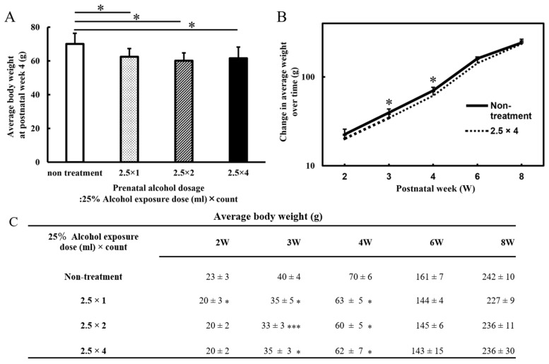
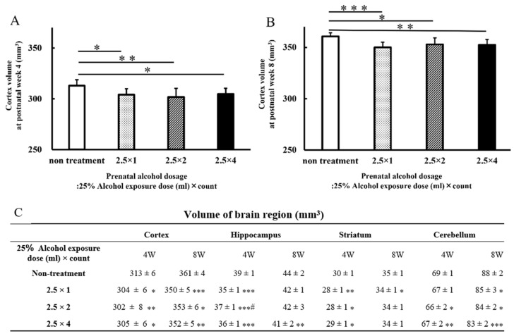
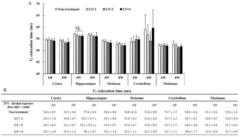
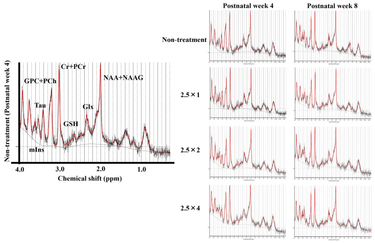
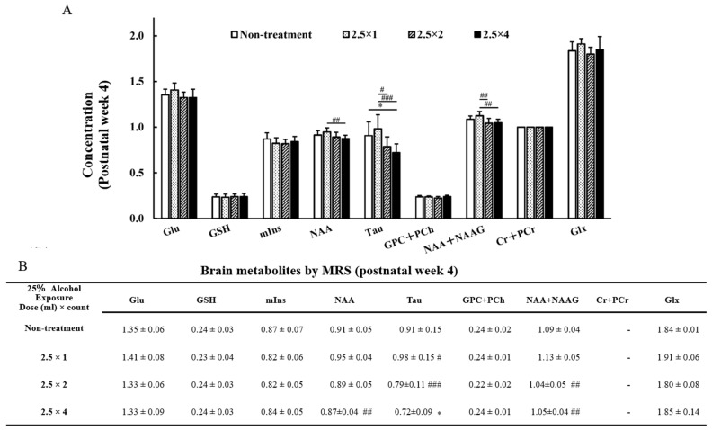
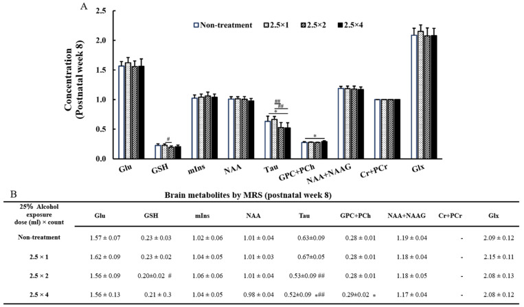
Prenatal alcohol exposure causes many detrimental alcohol-induced defects in children, collectively known as fetal alcohol spectrum disorders (FASD). This study aimed to evaluate a rat model of FASD, in which alcohol was administered at progressively increasing doses during late pregnancy, using preclinical magnetic resonance (MR) imaging (MRI) and MR spectroscopy (MRS). Wistar rats were orally administered 2.5 mL/day of ethanol (25% concentration) on gestational day 15, and postnatal fetuses were used as FASD models. Four groups were used: a control group (non-treatment group) and three groups of FASD model rats that received one, two, or four doses of ethanol, respectively, during the embryonic period. Body weight was measured every other week until eight weeks of age. MRI and MRS were performed at 4 and 8 weeks of age. The volume of each brain region was measured using acquired T-weighted images. At 4 weeks of age, body weight and cortex volume were significantly lower in the three FASD model groups (2.5 × 1: 304 ± 6 mm, < 0.05; 2.5 × 2: 302 ± 8 mm, < 0.01; 2.5 × 4: 305 ± 6 mm, < 0.05) than they were in the non-treatment group (non-treatment: 313 ± 6 mm). The FASD model group that received four doses of alcohol (2.5 × 4: 0.72 ± 0.09, < 0.05) had lower Taurine/Cr values than the non-treatment group did (non-treatment: 0.91 ± 0.15), an effect that continued at 8 weeks of age (non-treatment: 0.63 ± 0.09; 2.5 × 4: 0.52 ± 0.09, < 0.05). This study is the first to assess brain metabolites and volume over time using MRI and MRS. Decreases in brain volume and taurine levels were observed at 4 and 8 weeks of age, suggesting that the effects of alcohol persisted beyond adulthood.
产前酒精暴露会导致儿童出现许多由酒精引起的有害缺陷,统称为胎儿酒精谱系障碍(FASD)。本研究旨在利用临床前磁共振成像(MRI)和磁共振波谱(MRS)评估一种FASD大鼠模型,该模型在妊娠后期给予逐渐增加剂量的酒精。在妊娠第15天,对Wistar大鼠口服给予2.5 mL/天的乙醇(浓度为25%),产后胎儿用作FASD模型。实验分为四组:一个对照组(未处理组)和三组FASD模型大鼠,它们在胚胎期分别接受一剂、两剂或四剂乙醇。每隔一周测量一次体重,直至八周龄。在4周龄和8周龄时进行MRI和MRS检查。使用获取的T加权图像测量每个脑区的体积。在4周龄时,三个FASD模型组的体重和皮质体积(2.5×1:304±6 mm,P<0.05;2.5×2:302±8 mm,P<0.01;2.5×4:305±6 mm,P<0.05)显著低于未处理组(未处理:313±6 mm)。接受四剂酒精的FASD模型组(2.5×4:0.72±0.09,P<0.05)的牛磺酸/肌酸(Taurine/Cr)值低于未处理组(未处理:0.91±0.15),这种影响在8周龄时仍然存在(未处理:0.63±0.09;2.5×4:0.52±0.09,P<0.05)。本研究首次使用MRI和MRS随时间评估脑代谢物和体积。在4周龄和8周龄时观察到脑体积和牛磺酸水平降低,这表明酒精的影响持续到成年期以后。