Institute of Mental Health Research at the Royal, University of Ottawa, Ottawa, Ontario, Canada.
Translational Neuroimaging Laboratory, The McGill University Research Centre for Studies in Aging, McGill University, Montreal, Quebec, Canada.
Hum Brain Mapp. 2023 Jun 15;44(9):3913-3925. doi: 10.1002/hbm.26324. Epub 2023 May 1.
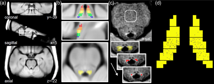
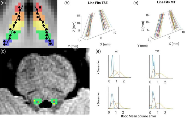
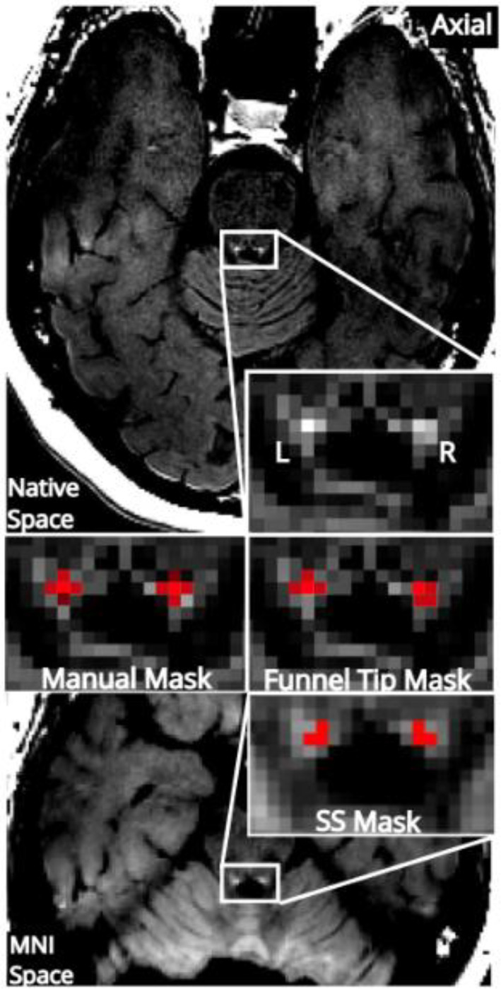
Following the development of magnetic resonance imaging (MRI) methods to assay the integrity of catecholamine nuclei, including the locus coeruleus (LC), there has been an effort to develop automated methods that can accurately segment this small structure in an automated manner to promote its widespread use and overcome limitations of manual segmentation. Here we characterize an automated LC segmentation approach (referred to as the funnel-tip [FT] method) in healthy individuals and individuals with LC degeneration in the context of Alzheimer's disease (AD, confirmed with tau-PET imaging using [18F]MK6240). The first sample included n = 190 individuals across the AD spectrum from cognitively normal to moderate AD. LC signal assayed with FT segmentation showed excellent agreement with manual segmentation (intraclass correlation coefficient [ICC] = 0.91). Compared to other methods, the FT method showed numerically higher correlation to AD status (defined by presence of tau: Cohen's d = 0.64) and AD severity (Braak stage: Pearson R = -.35, cognitive function: R = .25). In a separate sample of n = 12 control participants, the FT method showed excellent scan-rescan reliability (ICC = 0.82). In another sample of n = 30 control participants, we found that the structure of the LC defined by FT segmentation approximated its expected shape as a contiguous line: <5% of LC voxels strayed >1 voxel (0.69 mm) from this line. The FT LC segmentation shows high agreement with manual segmentation and captures LC degeneration in AD. This practical method may facilitate larger research studies of the human LC-norepinephrine system and has potential to support future use of neuromelanin-sensitive MRI as a clinical biomarker.
随着磁共振成像(MRI)方法的发展,用于检测儿茶酚胺核(包括蓝斑核(LC))的完整性,人们一直在努力开发能够自动准确分割这个小结构的自动方法,以促进其广泛应用并克服手动分割的局限性。在这里,我们在阿尔茨海默病(AD,使用 [18F]MK6240 进行 tau-PET 成像进行确认)背景下,对健康个体和 LC 退化个体描述了一种自动 LC 分割方法(称为漏斗尖端 [FT] 方法)。第一个样本包括 n = 190 名个体,涵盖了从认知正常到中度 AD 的 AD 谱。使用 FT 分割测量的 LC 信号与手动分割具有极好的一致性(组内相关系数 [ICC] = 0.91)。与其他方法相比,FT 方法与 AD 状态的相关性更高(根据 tau 的存在定义:Cohen 的 d = 0.64),与 AD 严重程度的相关性更高(Braak 分期:Pearson R = -.35,认知功能:R = -.25)。在另一个 n = 12 名对照参与者的样本中,FT 方法显示出极好的扫描重测可靠性(ICC = 0.82)。在另一个 n = 30 名对照参与者的样本中,我们发现 FT 分割定义的 LC 结构近似于其作为连续线的预期形状:<5%的 LC 体素偏离该线> 1 个体素(0.69 毫米)。FT LC 分割与手动分割具有高度一致性,并捕获了 AD 中的 LC 退化。这种实用方法可能有助于进行更大规模的人类 LC-去甲肾上腺素系统研究,并有可能支持未来使用神经黑色素敏感 MRI 作为临床生物标志物。