Department of Rheumatology, Ghent University Hospital, Ghent, Belgium
Molecular Immunology and Inflammation Unit, VIB-UGent Center for Inflammation Research, Zwijnaarde, Belgium.
RMD Open. 2023 May;9(2). doi: 10.1136/rmdopen-2023-002994.
To examine radiographic axial damage of the sacroiliac joints and spine in patients with psoriatic arthritis (PsA) and spondyloarthritis (SpA) in private and academic Belgian practices.
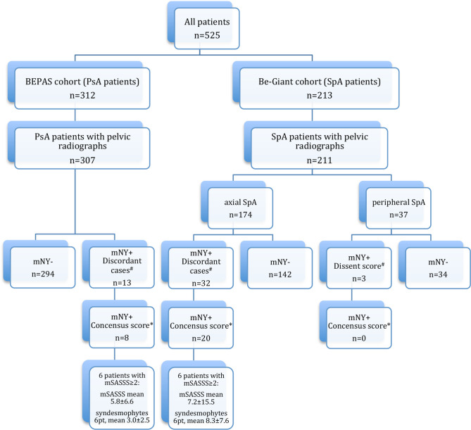
Patients with PsA with clinical diagnosis of PsA and fulfilling the Classification Criteria for Psoriatic Arthritis from the prospective Belgian Epidemiological Psoriatic Arthritis Study and patients with SpA fulfilling the Assessment of SpondyloArthritis international Society classification criteria for SpA originate from the Ghent and BelGian Inflammatory Arthritis and spoNdylitis cohorTs were included in this study. Baseline pelvic and spinal radiographs were analysed by two calibrated readers. Blinded for the origin of the cohort or clinical data readers assessed the modified Stoke Ankylosing Spondylitis Spine Score (mSASSS) and modified New York criteria on spinal and pelvic radiographs, respectively. Data were compared between both patient groups.
Of the 525 patients included (312 PsA and 213 SpA), most patients showed normal spinal radiographs: 87.5% of the patients with PsA and 92.0% of the patients with SpA. Patients with SpA with spinal damage show higher mSASSS than the patients with PsA (p<0.05). In patients with PsA, cervical spine is more often affected; 24/33 patients (72.7%) compared with lumbar spine 11/33 (33.3%). While in patients with SpA, syndesmophyte location was more evenly distributed; cervical 9/14 (64.3%) and lumbar 10/14 (71.4%).
Minimal radiographic spinal damage was observed in Belgian patients with PsA or SpA. Patients with SpA tend to have higher mSASSS values and more syndesmophytes compared with PsA. Syndesmophytes were more often located in the cervical spine of patients with PsA, while the location was equally distributed in axSpA.
在私人和学术的比利时诊所中,检查患有银屑病关节炎(PsA)和脊柱关节炎(SpA)的患者的骶髂关节和脊柱的放射轴向损伤。
本研究纳入了来自前瞻性比利时银屑病关节炎研究的具有临床诊断为 PsA 且符合银屑病关节炎分类标准的 PsA 患者,以及符合脊柱关节炎国际协会分类标准的 SpA 患者。基线骨盆和脊柱 X 线片由两名经过校准的读者进行分析。读者在不了解队列来源或临床数据的情况下,分别评估改良的 Stoke 强直性脊柱炎脊柱评分(mSASSS)和改良的纽约标准在脊柱和骨盆 X 线片上的表现。比较两组患者的数据。
在纳入的 525 例患者中(312 例 PsA 和 213 例 SpA),大多数患者的脊柱 X 线片正常:87.5%的 PsA 患者和 92.0%的 SpA 患者。有脊柱损伤的 SpA 患者的 mSASSS 高于 PsA 患者(p<0.05)。在 PsA 患者中,颈椎更常受累;33 例患者中有 24 例(72.7%),而腰椎受累仅 11 例(33.3%)。而在 SpA 患者中,骨桥的位置分布更为均匀;颈椎 9/14 例(64.3%)和腰椎 10/14 例(71.4%)。
在比利时的 PsA 或 SpA 患者中,观察到轻微的放射学脊柱损伤。与 PsA 相比,SpA 患者的 mSASSS 值更高,且骨桥更多。在 PsA 患者中,骨桥更常位于颈椎,而在 axSpA 中,骨桥的位置分布更为均匀。