Department of Biochemistry, Purdue University, West Lafayette, IN, 47907, USA.
Department of Medical Neurobiology, Institute for Medical Research Israel-Canada (IMRIC), Edmond and Lily Safra Center for Brain Sciences (ELSC), Faculty of Medicine, The Hebrew University, Jerusalem, 91120, Israel.
Redox Biol. 2023 Jul;63:102723. doi: 10.1016/j.redox.2023.102723. Epub 2023 Apr 27.
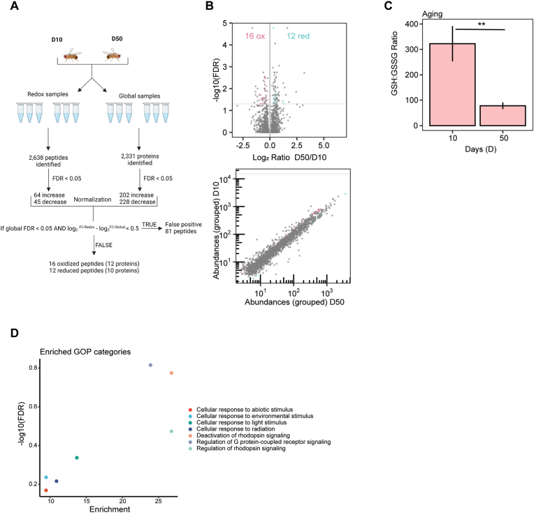
The retina is one of the highest oxygen-consuming tissues because visual transduction and light signaling processes require large amounts of ATP. Thus, because of the high energy demand, oxygen-rich environment, and tissue transparency, the eye is susceptible to excess production of reactive oxygen species (ROS) resulting in oxidative stress. Oxidative stress in the eye is associated with the development and progression of ocular diseases including cataracts, glaucoma, age-related macular degeneration, and diabetic retinopathy. ROS can modify and damage cellular proteins, but can also be involved in redox signaling. In particular, the thiol groups of cysteines can undergo reversible or irreversible oxidative post-translational modifications (PTMs). Identifying the redox-sensitive cysteines on a proteome-wide scale provides insight into those proteins that act as redox sensors or become irreversibly damaged upon exposure to oxidative stress. In this study, we profiled the redox proteome of the Drosophila eye under prolonged, high intensity blue light exposure and age using iodoacetamide isobaric label sixplex reagents (iodo-TMT) to identify changes in cysteine availability. Although redox metabolite analysis of the major antioxidant, glutathione, revealed similar ratios of its oxidized and reduced form in aged or light-stressed eyes, we observed different changes in the redox proteome under these conditions. Both conditions resulted in significant oxidation of proteins involved in phototransduction and photoreceptor maintenance but affected distinct targets and cysteine residues. Moreover, redox changes induced by blue light exposure were accompanied by a large reduction in light sensitivity that did not arise from a reduction in the photopigment level, suggesting that the redox-sensitive cysteines we identified in the phototransduction machinery might contribute to light adaptation. Our data provide a comprehensive description of the redox proteome of Drosophila eye tissue under light stress and aging and suggest how redox signaling might contribute to light adaptation in response to acute light stress.
视网膜是耗氧量最高的组织之一,因为视觉转导和光信号过程需要大量的 ATP。因此,由于高能量需求、富含氧气的环境和组织透明度,眼睛容易产生过多的活性氧物种 (ROS),导致氧化应激。眼睛中的氧化应激与包括白内障、青光眼、年龄相关性黄斑变性和糖尿病性视网膜病变在内的眼部疾病的发展和进展有关。ROS 可以修饰和破坏细胞蛋白,但也可以参与氧化还原信号。特别是,半胱氨酸的巯基可以经历可逆或不可逆的氧化翻译后修饰 (PTM)。在全蛋白质组范围内鉴定氧化还原敏感的半胱氨酸,可深入了解那些作为氧化还原传感器的蛋白质,或在暴露于氧化应激时变得不可逆损伤的蛋白质。在这项研究中,我们使用碘乙酰胺等重六plex 试剂 (碘-TMT) 对果蝇眼睛进行了长时间、高强度蓝光照射和年龄的氧化还原蛋白质组分析,以确定半胱氨酸可用性的变化。尽管主要抗氧化剂谷胱甘肽的氧化还原代谢物分析显示其氧化和还原形式在衰老或光应激眼中的比例相似,但我们观察到这些条件下氧化还原蛋白质组的不同变化。这两种情况都导致光转导和光感受器维持所涉及的蛋白质发生显著氧化,但影响了不同的靶标和半胱氨酸残基。此外,蓝光照射引起的氧化还原变化伴随着光敏感性的大幅降低,而这种降低并非源于光色素水平的降低,这表明我们在光转导机制中鉴定的氧化还原敏感半胱氨酸可能有助于光适应。我们的数据提供了光应激和衰老下果蝇眼组织氧化还原蛋白质组的全面描述,并提出了氧化还原信号如何有助于对急性光应激的光适应。