Laboratory of Experimental Psychiatry, Instituto Nacional de Neurología y Neurocirugía, Mexico City, Mexico; Neuropsychiatry Department, Instituto Nacional de Neurología y Neurocirugía, Mexico City, Mexico.
Neuropsychiatry Department, Instituto Nacional de Neurología y Neurocirugía, Mexico City, Mexico.
Brain Behav Immun. 2023 Jul;111:270-276. doi: 10.1016/j.bbi.2023.05.001. Epub 2023 May 5.
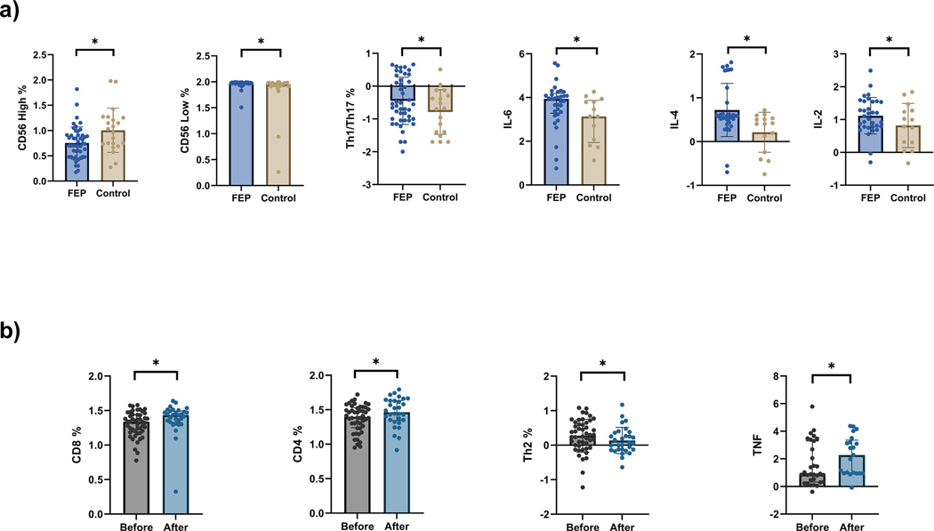
Studies of cellular and cytokine profiles have contributed to the inflammation hypothesis of schizophrenia; however, precise markers of inflammatory dysfunction remain elusive. A number of proton magnetic resonance spectroscopy (1H-MRS) studies in patients with first-episode psychosis (FEP) have shown higher brain levels of metabolites such as glutamate, myo-inositol (mI) and choline-containing compounds (tCho), suggesting neuroinflammation. Here, we present peripheral inflammatory profiles in antipsychotic-naive FEP patients and age-and-sex matched healthy controls, as well as cortical glutamate, mI and tCho levels using 1H-MRS. Inflammatory profiles were analyzed using cytokine production by peripheral blood mononuclear cells, that were either spontaneous or stimulated, in 48 FEP patients and 23 controls. 1H-MRS of the medial prefrontal cortex was obtained in 29 FEP patients and 18 controls. Finally, 16 FEP patients were rescanned after 4 weeks of treatment (open-label) with Risperidone. FEP patients showed a higher proportion of proinflammatory Th1/Th17 subset, and an increased spontaneous production of Interleukin (IL)-6, IL-2 and IL-4 compared with the control group. Results obtained from 1H-MRS showed no significant difference in either glutamate, mI or tCho between FEP and control groups. At baseline, CD8% showed a negative correlation with glutamate in FEP patients; after 4 weeks of risperidone treatment, the FEP group exhibited a decrease in glutamate levels which positively correlated with CD4 + T cells. Nevertheless, these correlations did not survive correction for multiple comparisons. FEP patients show evidence of immune dysregulation, affecting both the innate and adaptive immune response, with a predominantly Th2 signature. These findings, along with the changes produced by antipsychotic treatment, could be associated with both systemic and central inflammatory processes in schizophrenia.
细胞和细胞因子谱的研究促进了精神分裂症炎症假说的发展;然而,确切的炎症功能障碍标志物仍然难以捉摸。许多首发精神病(FEP)患者的质子磁共振波谱(1H-MRS)研究表明,谷氨酸、肌醇(mI)和含胆碱化合物(tCho)等代谢物的脑水平升高,提示存在神经炎症。在这里,我们介绍了未经抗精神病药物治疗的 FEP 患者和年龄及性别匹配的健康对照者的外周炎症谱,以及使用 1H-MRS 检测的皮质谷氨酸、mI 和 tCho 水平。通过 48 名 FEP 患者和 23 名对照者的外周血单个核细胞自发或刺激后的细胞因子产生情况分析了炎症谱。对 29 名 FEP 患者和 18 名对照者进行了内侧前额叶皮质的 1H-MRS 检查。最后,16 名 FEP 患者在利培酮(开放性标签)治疗 4 周后进行了重新扫描。与对照组相比,FEP 患者表现出更高比例的促炎 Th1/Th17 亚群,以及更高的 IL-6、IL-2 和 IL-4 自发产生。1H-MRS 结果显示 FEP 组和对照组之间谷氨酸、mI 或 tCho 无显著差异。在基线时,FEP 患者的 CD8%与谷氨酸呈负相关;在利培酮治疗 4 周后,FEP 组的谷氨酸水平下降,与 CD4+T 细胞呈正相关。然而,这些相关性在经过多次比较校正后并不成立。FEP 患者表现出免疫失调的证据,影响先天和适应性免疫反应,以 Th2 表型为主。这些发现,以及抗精神病药物治疗产生的变化,可能与精神分裂症的全身和中枢炎症过程有关。