Departments of Pathology Seoul National University Hospital, Seoul National University College of Medicine, Seoul, Korea.
Departments of Radiology, Seoul National University Hospital, Seoul National University College of Medicine, Seoul, Korea.
Clin Mol Hepatol. 2023 Jul;29(3):733-746. doi: 10.3350/cmh.2023.0034. Epub 2023 May 8.
BACKGROUND/AIMS: The microvascular invasion (MVI) of hepatocellular carcinoma (HCC) involves a wide histological spectrum, and it is unclear whether the degree of MVI correlates with patient prognosis or imaging findings. Here, we evaluate the prognostic value of MVI classification and analyze the radiologic features predictive of MVI.
Using a retrospective cohort of 506 patients with resected solitary HCCs, the histological and imaging features of MVI were reviewed and correlated with clinical data.
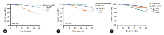
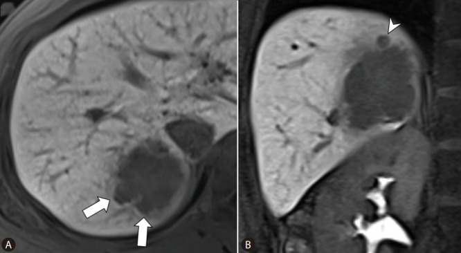
MVI-positive HCCs invading ≥5 vessels or those with ≥50 invaded tumor cells were significantly associated with decreased overall survival (OS). The 5-year OS, recurrence-free survival (RFS), and beyond Milan criteria RFS rates were significantly poorer in patients with severe MVI compared with those with mild or no MVI. Severe MVI was a significant independent predictive factor for OS (odds ratio [OR], 2.962; p<0.001), RFS (OR, 1.638; p=0.002), and beyond Milan criteria RFS (OR, 2.797; p<0.001) on multivariable analysis. On MRI, non-smooth tumor margins (OR, 2.224; p=0.023) and satellite nodules (OR, 3.264; p<0.001) were independently associated with the severe-MVI group on multivariable analysis. Both non-smooth tumor margins and satellite nodules were associated with worse 5-year OS, RFS, and beyond Milan criteria RFS.
Histologic risk classification of MVI according to the number of invaded microvessels and invading carcinoma cells was a valuable predictor of prognosis in HCC patients. Non-smooth tumor margin and satellite nodules were significantly associated with severe MVI and poor prognosis.
背景/目的:肝细胞癌(HCC)的微血管侵犯(MVI)涉及广泛的组织学谱,并且尚不清楚 MVI 的程度是否与患者预后或影像学表现相关。在这里,我们评估 MVI 分类的预后价值,并分析预测 MVI 的影像学特征。
使用 506 例接受根治性单发 HCC 切除术患者的回顾性队列,回顾性分析 MVI 的组织学和影像学特征,并与临床数据相关联。
侵犯≥5 支血管或≥50 个侵袭性肿瘤细胞的 MVI 阳性 HCC 患者的总生存期(OS)明显降低。与轻度或无 MVI 的患者相比,严重 MVI 的患者 5 年 OS、无复发生存率(RFS)和超过米兰标准的 RFS 率明显较差。严重 MVI 是 OS(优势比 [OR],2.962;p<0.001)、RFS(OR,1.638;p=0.002)和超过米兰标准的 RFS(OR,2.797;p<0.001)的独立预测因素。在 MRI 上,非平滑肿瘤边缘(OR,2.224;p=0.023)和卫星结节(OR,3.264;p<0.001)在多变量分析中与严重-MVI 组独立相关。非平滑肿瘤边缘和卫星结节均与较差的 5 年 OS、RFS 和超过米兰标准的 RFS 相关。
根据侵犯微血管的数量和侵袭性癌细胞对 MVI 进行组织学风险分类是 HCC 患者预后的有价值的预测指标。非平滑肿瘤边缘和卫星结节与严重 MVI 和不良预后显著相关。