Heller Gordon D
Department of Radiology, Icahn School of Medicine, Division of Neuroradiology-West, 1000 10th Avenue, New York, NY 10009, USA.
Ther Adv Rare Dis. 2020 Oct 29;1:2633004020969702. doi: 10.1177/2633004020969702. eCollection 2020 Jan-Dec.
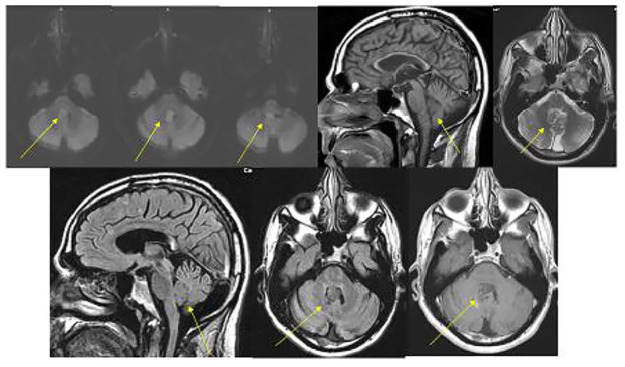
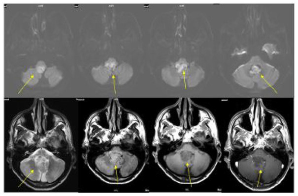
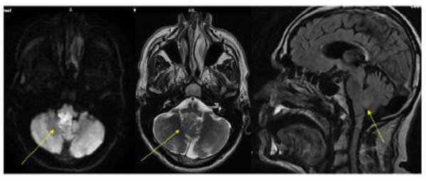
Gardner Syndrome is a rare disease with clinical manifestations of familial intestinal polyposis with osteomas. Cutaneous and subcutaneous lesions are common and epidermoid cyst is a characteristic dermatologic finding. This case report presents a novel finding of an intracranial epidermoid situated in the fourth ventricle in a patient with Gardner Syndrome. This intracranial epidermoid has been followed with sequential magnetic resonance imaging (MRI) for 10 years with progressive growth of the lesion. This suggests the conservative management is an option in patients with an enlarging epidermoid cyst in the fourth ventricle.
Gardner syndrome is a rare disease and form of familial adenomatous polyposis (FAP) that is characterized by multiple small growths of cells (polyps) in the colon and various types of tumors, both noncancerous (benign) and cancerous (malignant). It is caused by changes (mutations) in the APC gene. Abnormal changes on the skin and under the skin are common as well as growths called epidermoid cysts. The cysts develop when cells that are meant to become skin, hair, and nails (epithelial cells) are trapped among the cells that form the brain. Epidermoid brain cysts may be diagnosed by magnetic resonance imaging (MRI) and computerized tomography (CT) scans. Typical treatment usually involves surgery. To present a different management strategy for patients with Gardner Syndrome with epidermoid brain cysts. This patient is the first patient with Gardner Syndrome with a very rare epidermoid brain cyst reported to be treated in a conservative manner.The patient was monitored for 10 years with regular MRI scans and the cyst continued to grow over this time.Despite this growth the patient has shown no signs of a buildup of fluid in the cavities deep within the brain (called hydrocephalus).The patient experienced nonfocal headaches, which were relieved with medication so doctors decided not to surgically remove the cyst. Conservative management of epidermoid brain cysts in Gardner patients has not been reported before. This case report shows that conservative management may be an alternative option for patients with a growing epidermoid cyst in the fourth ventricle of the brain. Conservative treatment is designed to avoid invasive treatments or surgery and provides a different option for patients who are unable to have surgery.
加德纳综合征是一种罕见疾病,临床表现为伴有骨瘤的家族性肠息肉病。皮肤和皮下病变很常见,表皮样囊肿是其特征性的皮肤表现。本病例报告展示了一名加德纳综合征患者第四脑室内存在颅内表皮样囊肿的新发现。该颅内表皮样囊肿已通过连续磁共振成像(MRI)随访10年,病变呈进行性生长。这表明对于第四脑室内表皮样囊肿增大的患者,保守治疗是一种选择。
加德纳综合征是一种罕见疾病,也是家族性腺瘤性息肉病(FAP)的一种形式,其特征是结肠内有多个小细胞生长物(息肉)以及各种类型的肿瘤,包括非癌性(良性)和癌性(恶性)肿瘤。它由APC基因突变引起。皮肤和皮下的异常变化以及表皮样囊肿很常见。当本应形成皮肤、毛发和指甲的细胞(上皮细胞)被困在构成大脑的细胞之间时,就会形成囊肿。表皮样脑囊肿可通过磁共振成像(MRI)和计算机断层扫描(CT)进行诊断。典型的治疗通常包括手术。 为患有表皮样脑囊肿的加德纳综合征患者提供一种不同的治疗策略。 该患者是首例报道采用保守治疗的患有非常罕见的表皮样脑囊肿的加德纳综合征患者。该患者通过定期MRI扫描监测了10年,在此期间囊肿持续生长。尽管囊肿生长,但患者未出现脑深部腔隙内积液增多的迹象(称为脑积水)。患者经历了非局灶性头痛,药物治疗后缓解,因此医生决定不通过手术切除囊肿。此前尚未报道过对加德纳综合征患者的表皮样脑囊肿进行保守治疗。本病例报告表明,对于脑第四脑室内表皮样囊肿不断增大的患者,保守治疗可能是一种替代选择。保守治疗旨在避免侵入性治疗或手术,为无法进行手术的患者提供了不同的选择。