Beyer Margaret, France John, Nagaraja Tavarekere N, Lavik Erin B, Knight Robert A, Lewandowski Christopher A, Miller Joseph B
Department of Emergency Medicine, Henry Ford Hospital, Detroit, MI, USA.
Department of Psychiatry and Behavioral Neurosciences, Wayne State University, Detroit, MI, USA.
Brain Circ. 2022 Dec 6;8(4):228-231. doi: 10.4103/bc.bc_45_22. eCollection 2022 Oct-Dec.
Hemostatic nanoparticles (hNPs) have shown efficacy in decreasing intracerebral hemorrhage (ICH) in animal models and are suggested to be of use to counter tissue plasminogen activator (tPA)-induced acute ICH.
The objective of this study was to test the ability of an hNP preparation to alter the clotting properties of blood exposed to tPA .
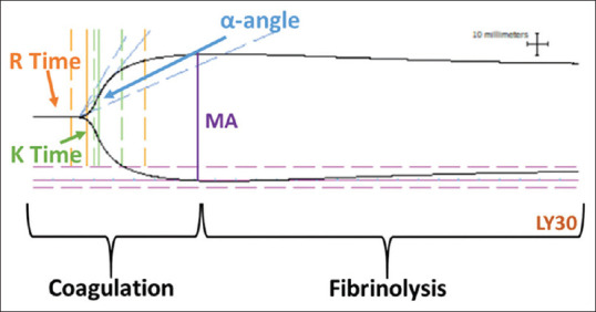
Fresh blood samples were obtained from normal male Sprague-Dawley rats (~300 g; = 6) and prepared for coagulation assays by thromboelastography (TEG) methods. Samples were untreated, exposed to tPA, or exposed to tPA and then to hNP. TEG parameters included reaction time (R, time in minutes elapsed from test initiation to initial fibrin formation), coagulation time (K, time in minutes from R until initial clot formation), angle (α, a measure in degrees of the rate of clot formation), maximum amplitude (MA, the point when the clot reaches its MA in mm), lysis at 30 min after MA (LY30, %), and clot strength (G, dynes/cm), an index of clot strength.
Kruskal-Wallis test was employed to compare TEG parameters measured for untreated control samples versus those exposed to tPA and to compare tPA-exposed samples to samples treated with tPA + hNPs. Significances were inferred at ≤ 0.05.
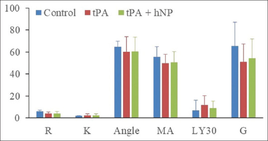
Compared to untreated samples, tPA-treated samples showed a trend toward decreased angle and G suggesting potentially clot formation rate and clot strength. The addition of hNP did not affect any of these or other measured indices.
The data demonstrated no hemostatic effects when the hNP was used in the presence of tPA. The lack of change in any of the TEG parameters measured in the present study may indicate limitations of the hNPs to reverse the thrombolytic cascade initiated by tPA.
止血纳米颗粒(hNP)在动物模型中已显示出减少脑出血(ICH)的效果,并被认为可用于对抗组织型纤溶酶原激活剂(tPA)诱导的急性脑出血。
本研究的目的是测试一种hNP制剂改变暴露于tPA的血液凝血特性的能力。
从正常雄性Sprague-Dawley大鼠(约300g;n = 6)获取新鲜血液样本,并通过血栓弹力图(TEG)方法准备用于凝血试验。样本未处理、暴露于tPA或暴露于tPA后再暴露于hNP。TEG参数包括反应时间(R,从测试开始到初始纤维蛋白形成所经过的分钟数)、凝血时间(K,从R到初始凝块形成的分钟数)、角度(α,凝块形成速率的度数测量值)、最大振幅(MA,凝块达到其最大毫米振幅时的点)、MA后30分钟的溶解率(LY30,%)和凝块强度(G,达因/厘米),凝块强度指标。
采用Kruskal-Wallis检验比较未处理对照样本与暴露于tPA的样本所测量的TEG参数,并比较暴露于tPA的样本与用tPA + hNP处理的样本。显著性推断为p≤0.05。
与未处理样本相比,tPA处理的样本显示角度和G有降低趋势,提示潜在的凝块形成速率和凝块强度降低。添加hNP并未影响这些或其他测量指标中的任何一项。
数据表明在tPA存在的情况下使用hNP没有止血作用。本研究中测量的任何TEG参数均未发生变化,这可能表明hNP在逆转由tPA引发的溶栓级联反应方面存在局限性。