Benabdallah Nadia, Bernardini Michela, Bianciardi Marta, de Labriolle-Vaylet Claire, Franck Didier, Desbrée Aurélie
Internal Dose Assessment Laboratory, Institute for Radiological Protection and Nuclear Safety (IRSN), Fontenay-aux-Roses, France.
Nuclear Medicine Department, European Hospital George Pompidou (HEGP), Paris, France.
EJNMMI Res. 2019 Feb 21;9(1):20. doi: 10.1186/s13550-019-0488-7.
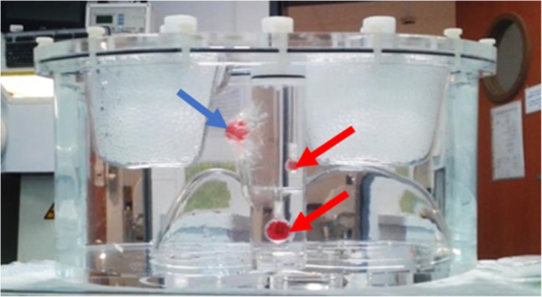
Ra imaging is crucial to evaluate the successfulness of the therapy of bone metastasis of castration-resistant prostate cancer (CRPC). The goals of this study were to establish a quantitative tomographic Ra imaging protocol with clinically achievable conditions, as well as to investigate its usefulness and limitations. We performed several experiments using the Infinia Hawkeye 4 gamma camera (GE) and physical phantoms in order to assess the optimal image acquisition and reconstruction parameters, such as the windows setting, as well as the iteration number and filter of the reconstruction algorithm. Then, based on the MIRD pamphlet 23, we used a NEMA phantom and an anthropomorphic TORSO® phantom to calibrate the gamma camera and investigate the accuracy of quantification.
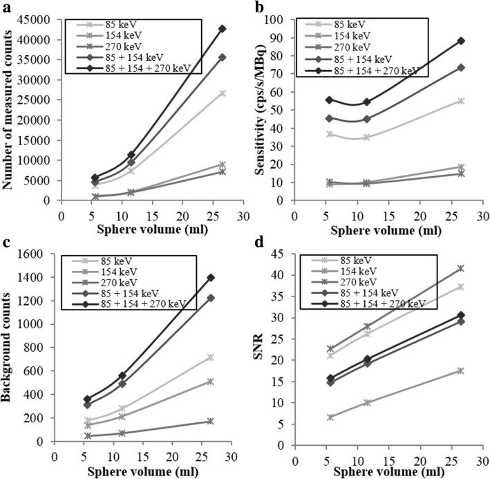
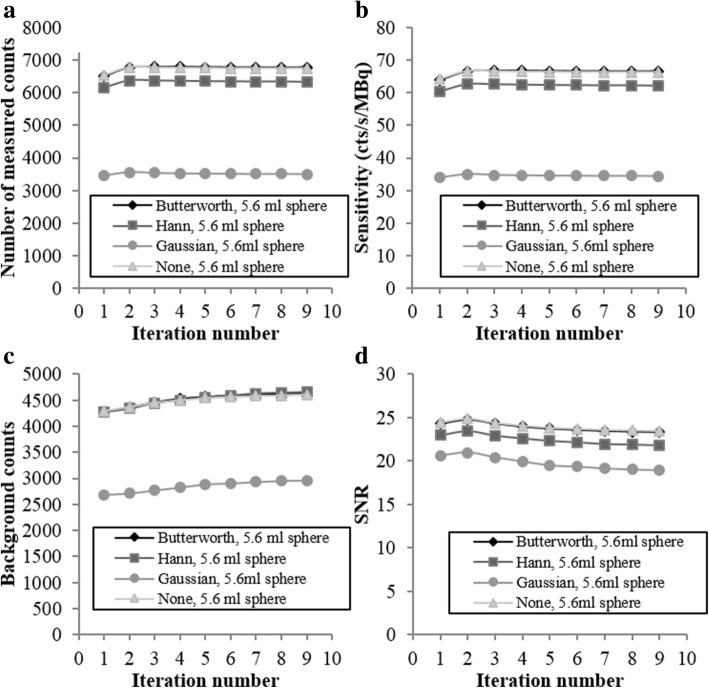
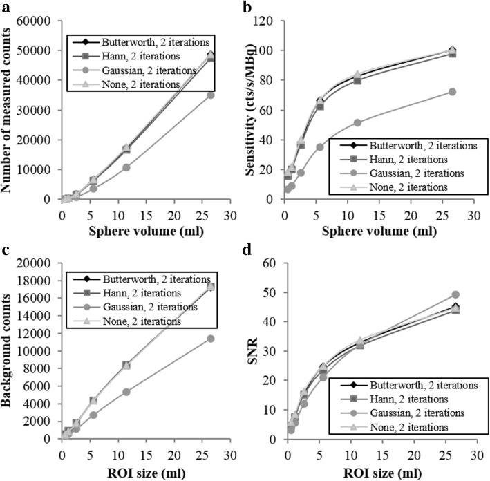
Experiences showed that the 85 keV ± 20%, 154 keV ± 10%, and 270 keV ± 10% energy windows are the most suitable for Ra imaging. The study with the NEMA phantom showed that the OSEM algorithm with 2 iterations, 10 subsets, and the Butterworth filter offered the best compromise between contrast and noise. Moreover, the calibration factors for different sphere sizes (26.5 ml, 11.5 ml, and 5.6 ml) were constant for Ra concentrations ranging between 6.5 and 22.8 kBq/ml. The values found are 73.7 cts/s/MBq, 43.8 cts/s/MBq, and 43.4 cts/s/MBq for 26.5 ml, 11.5 ml, and 5.6 ml sphere, respectively. For concentration lower than 6.5 kBq/ml, the calibration factors exhibited greater variability pointing out the limitations of SPECT/CT imaging for quantification. By the use of a TORSO® phantom, we simulated several tumors to normal tissue ratios as close as possible to clinical conditions. Using the calibration factors obtained with the NEMA phantom, for Ra concentrations higher than 8 kBq/ml, we were able to quantify the activity with an error inferior to 18.8% in a 5.6 ml lesion.
Absolute quantitative Ra SPECT imaging appears feasible once the dimension of the target is determined. Further evaluation should be needed to apply the calibration factor-based quantitation to clinical Ra SPECT/CT imaging. This will open the possibility for patient-specific Ra treatment planning and therapeutic outcome prediction in patients.
镭成像对于评估去势抵抗性前列腺癌(CRPC)骨转移治疗的成功与否至关重要。本研究的目的是建立一种在临床可实现条件下的定量断层镭成像方案,并研究其有效性和局限性。我们使用Infinia Hawkeye 4伽马相机(GE)和物理模型进行了多项实验,以评估最佳图像采集和重建参数,如窗宽设置以及重建算法的迭代次数和滤波器。然后,基于MIRD手册23,我们使用NEMA模型和拟人化TORSO®模型对伽马相机进行校准,并研究定量的准确性。
经验表明,85 keV±20%、154 keV±10%和270 keV±10%的能量窗最适合镭成像。使用NEMA模型的研究表明,采用2次迭代、10个子集和巴特沃斯滤波器的OSEM算法在对比度和噪声之间提供了最佳折衷。此外,对于浓度在6.5至22.8 kBq/ml之间的镭,不同球体尺寸(26.5 ml、11.5 ml和5.6 ml)的校准因子是恒定的。对于26.5 ml、11.5 ml和5.6 ml的球体,分别得到的值为73.7 cts/s/MBq、43.8 cts/s/MBq和43.4 cts/s/MBq。对于浓度低于6.5 kBq/ml的情况,校准因子表现出更大的变异性,指出了SPECT/CT成像在定量方面的局限性。通过使用TORSO®模型,我们模拟了几种尽可能接近临床情况的肿瘤与正常组织的比率。使用通过NEMA模型获得的校准因子,对于浓度高于8 kBq/ml的镭,我们能够在一个5.6 ml病变中以低于18.8%的误差对活性进行定量。
一旦确定了目标的尺寸,绝对定量镭SPECT成像似乎是可行的。需要进一步评估以将基于校准因子的定量应用于临床镭SPECT/CT成像。这将为患者特异性镭治疗计划和患者治疗结果预测开辟可能性。