Department of Radiology, Lausanne University Hospital (CHUV) and University of Lausanne (UNIL), Lausanne, Switzerland.
Department of Cardiovascular Imaging, Hôpital Cardiologique du Haut-Lévêque, CHU de Bordeaux, France.
Eur Radiol Exp. 2023 May 22;7(1):25. doi: 10.1186/s41747-023-00339-8.
To develop an isotropic three-dimensional (3D) T2 mapping technique for the quantitative assessment of the composition of knee cartilage with high accuracy and precision.
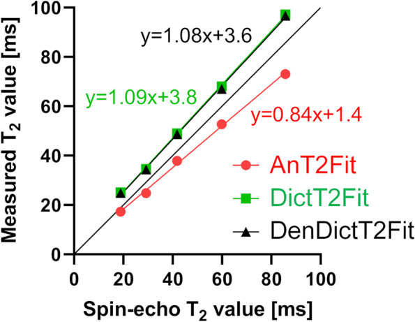
A T2-prepared water-selective isotropic 3D gradient-echo pulse sequence was used to generate four images at 3 T. These were used for three T2 map reconstructions: standard images with an analytical T2 fit (AnT2Fit); standard images with a dictionary-based T2 fit (DictT2Fit); and patch-based-denoised images with a dictionary-based T2 fit (DenDictT2Fit). The accuracy of the three techniques was first optimized in a phantom study against spin-echo imaging, after which knee cartilage T2 values and coefficients of variation (CoV) were assessed in ten subjects in order to establish accuracy and precision in vivo. Data given as mean ± standard deviation.
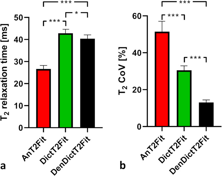
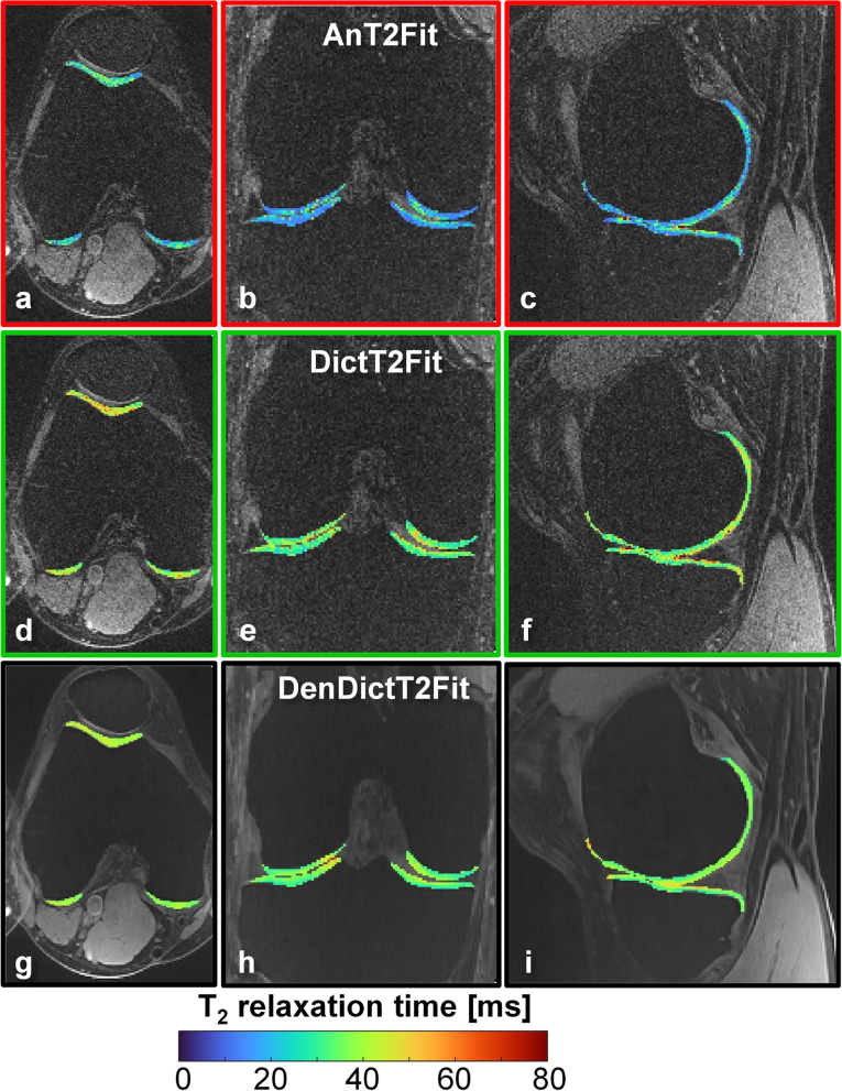
After optimization in the phantom, whole-knee cartilage T2 values of the healthy volunteers were 26.6 ± 1.6 ms (AnT2Fit), 42.8 ± 1.8 ms (DictT2Fit, p < 0.001 versus AnT2Fit), and 40.4 ± 1.7 ms (DenDictT2Fit, p = 0.009 versus DictT2Fit). The whole-knee T2 CoV reduced from 51.5% ± 5.6% to 30.5 ± 2.4 and finally to 13.1 ± 1.3%, respectively (p < 0.001 between all). The DictT2Fit improved the data reconstruction time: 48.7 ± 11.3 min (AnT2Fit) versus 7.3 ± 0.7 min (DictT2Fit, p < 0.001). Very small focal lesions were observed in maps generated with DenDictT2Fit.
Improved accuracy and precision for isotropic 3D T2 mapping of knee cartilage were demonstrated by using patch-based image denoising and dictionary-based reconstruction.
• Dictionary T2 fitting improves the accuracy of three-dimensional (3D) knee T2 mapping. • Patch-based denoising results in high precision in 3D knee T2 mapping. • Isotropic 3D knee T2 mapping enables the visualization of small anatomical details.
开发一种各向同性三维(3D)T2 映射技术,以高精度和高精准度定量评估膝关节软骨的成分。
在 3T 下使用 T2 预制备的水选择各向同性 3D 梯度回波脉冲序列生成四张图像。这些图像用于三种 T2 图谱重建:具有分析 T2 拟合的标准图像(AnT2Fit);基于字典的 T2 拟合的标准图像(DictT2Fit);以及基于字典的 T2 拟合的基于补丁的去噪图像(DenDictT2Fit)。首先在体模研究中针对自旋回波成像对这三种技术的准确性进行了优化,然后在十名受试者中评估了膝关节软骨 T2 值和变异系数(CoV),以确定体内的准确性和精密度。数据表示为平均值±标准差。
在体模优化后,健康志愿者的全膝关节软骨 T2 值为 26.6±1.6ms(AnT2Fit)、42.8±1.8ms(DictT2Fit,p<0.001 与 AnT2Fit 相比)和 40.4±1.7ms(DenDictT2Fit,p=0.009 与 DictT2Fit 相比)。全膝关节 T2 CoV 分别从 51.5%±5.6%降低到 30.5%±2.4%和 13.1%±1.3%(所有之间 p<0.001)。DictT2Fit 缩短了数据重建时间:48.7±11.3min(AnT2Fit)与 7.3±0.7min(DictT2Fit,p<0.001)。在使用基于补丁的图像去噪和基于字典的重建的 DenDictT2Fit 生成的图中观察到非常小的局灶性病变。
通过使用基于补丁的图像去噪和基于字典的重建,证明了各向同性 3D 膝关节软骨 T2 映射的准确性和精密度得到了提高。
字典 T2 拟合可提高三维(3D)膝关节 T2 映射的准确性。
基于补丁的去噪可实现 3D 膝关节 T2 映射的高精度。
各向同性 3D 膝关节 T2 映射可使小解剖细节可视化。