Suppr超能文献

3T 下用于半固态短 T 组织体内双指数弛豫测绘的 3D-T 准备零回波时间基于 PETRA 的序列。

3D-T prepared zero echo time-based PETRA sequence for in vivo biexponential relaxation mapping of semisolid short-T tissues at 3 T.

机构信息

Bernard and Irene Schwartz Center for Biomedical Imaging, Department of Radiology, New York University School of Medicine, New York, New York, USA.

出版信息

J Magn Reson Imaging. 2019 Oct;50(4):1207-1218. doi: 10.1002/jmri.26664. Epub 2019 Jan 28.

Abstract

BACKGROUND

In addition to the articular cartilage, osteoarthritis (OA) affects several other tissues such as tendons, ligaments, and subchondral bone. T1 relaxation study of these short T tissues may provide a more comprehensive evaluation of OA.

PURPOSE

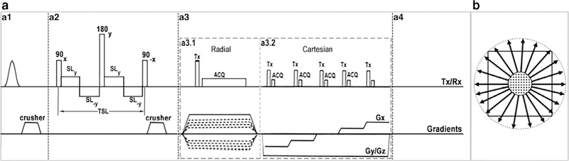
To develop a 3D spin-lattice relaxation in the rotating frame (T ) prepared zero echo time (ZTE)-based pointwise encoding time reduction with radial acquisition (3D-T -PETRA) sequence for relaxation mapping of semisolid short-T tissues on a clinical 3 T scanner.

STUDY TYPE

Prospective.

POPULATION

Phantom, two bovine whole knee joint and Achilles tendon specimens, 10 healthy volunteers with no known inflammation, trauma or pain in the knee or ankle.

FIELD STRENGTH/SEQUENCE: A customized PETRA sequence to acquire fat-suppressed 3D T -weighted images tissues with semisolid short T / relaxation times in the knee and ankle joints at 3 T.

ASSESSMENT

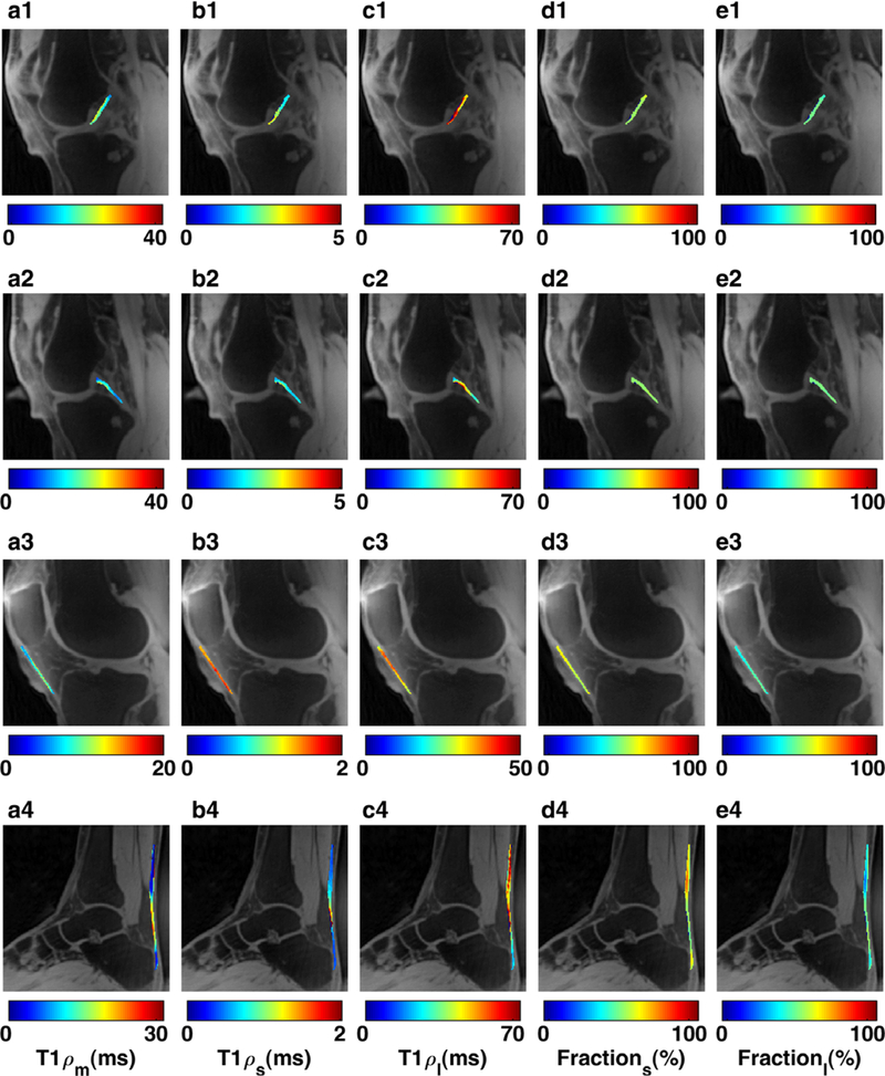
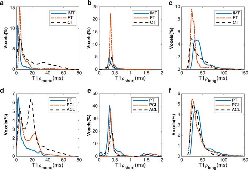
Mono- and biexponential T relaxation components were assessed in the patellar tendon (PT), anterior cruciate ligament (ACL), posterior cruciate ligament (PCL), and Achilles tendon (AT).

STATISTICAL TESTS

Kruskal-Wallis with post-hoc Dunn's test for multiple pairwise comparisons.

RESULTS

Phantom and ex vivo studies showed the feasibility of T relaxation mapping using the proposed 3D-T -PETRA sequence. The in vivo study demonstrated an averaged mono-T relaxation of (median [IQR]) 15.9 [14.5] msec, 23.6 [9.4] msec, 17.4 [7.4] msec, and 5.8 [10.2] msec in the PT, ACL, PCL, and AT, respectively. The bicomponent analysis showed the short and long components (with their relative fractions) of 0.65 [1.0] msec (46.9 [15.3]%) and 37.3 [18.4] msec (53.1 [15.3]%) for PT, 1.7 [2.1] msec (42.5 [12.5]%) and 43.7 [17.8] msec (57.5 [12.5]%) for ACL, and 1.2 [1.9] msec (42.6 [14.0]%) and 27.7 [14.7] msec (57.3 [14.0]%) for PCL and 0.4 [0.02] msec (58.8 [13.3]%/) and 31.3 [10.8] msec (41.2 [13.3]%) for AT. Statistically significant (P ≤ 0.05) differences were observed in the mono- and biexponential relaxation between several regions.

DATA CONCLUSION

The 3D-T -PETRA sequence allows volumetric, isotropic (0.78 × 0.78 × 0.78 mm), biexponential T assessment with corresponding fractions of the tissues with semisolid short T / .

LEVEL OF EVIDENCE

2 Technical Efficacy Stage: 1 J. Magn. Reson. Imaging 2019;50:1207-1218.

摘要

背景

除了关节软骨,骨关节炎(OA)还会影响到其他一些组织,如肌腱、韧带和软骨下骨。这些短 T 组织的 T1 弛豫研究可能为 OA 提供更全面的评估。

目的

在临床 3T 扫描仪上开发一种基于三维旋转晶格弛豫在旋转框架(T1)的准备零回波时间(ZTE)的点编码时间减少的径向采集(3D-T1-PETRA)序列,用于半固态短 T 组织的弛豫图。

研究类型

前瞻性。

人群

幻影、两个牛整个膝关节和跟腱标本、10 名无膝关节或踝关节炎症、创伤或疼痛的健康志愿者。

场强/序列:一种定制的 PETRA 序列,用于在 3T 时采集具有半固态短 T1/弛豫时间的膝关节和踝关节的脂肪抑制 3D T1 加权图像组织。

评估

在髌腱(PT)、前交叉韧带(ACL)、后交叉韧带(PCL)和跟腱(AT)中评估单指数和双指数 T1 弛豫分量。

统计检验

Kruskal-Wallis 检验,用于多个配对比较的事后 Dunn 检验。

结果

幻影和离体研究表明,使用所提出的 3D-T1-PETRA 序列进行 T1 弛豫映射是可行的。在体内研究中,在 PT、ACL、PCL 和 AT 中分别平均观察到单 T1 弛豫时间为(中位数[IQR])15.9[14.5]msec、23.6[9.4]msec、17.4[7.4]msec 和 5.8[10.2]msec。双指数分析显示,0.65[1.0]msec(46.9[15.3]%)和 37.3[18.4]msec(53.1[15.3]%)的短和长分量(相对分数)和 1.7[2.1]msec(42.5[12.5]%)和 43.7[17.8]msec(57.5[12.5]%)在 ACL 中,和 1.2[1.9]msec(42.6[14.0]%)和 27.7[14.7]msec(57.3[14.0]%)在 PCL 和 0.4[0.02]msec(58.8[13.3]%/)和 31.3[10.8]msec(41.2[13.3]%)在 AT 中。在几个区域的单指数和双指数弛豫之间观察到统计学显著(P≤0.05)差异。

数据结论

3D-T1-PETRA 序列允许具有半固态短 T1/弛豫时间的组织的体积、各向同性(0.78×0.78×0.78mm)、双指数 T1 评估以及相应的分数。

证据水平

2 技术功效阶段:1 J. Magn. Reson. Imaging 2019;50:1207-1218.

相似文献

1
3D-T prepared zero echo time-based PETRA sequence for in vivo biexponential relaxation mapping of semisolid short-T tissues at 3 T.
J Magn Reson Imaging. 2019 Oct;50(4):1207-1218. doi: 10.1002/jmri.26664. Epub 2019 Jan 28.
2
Age and Gender-Dependence of Single-and Bi-Exponential T MR Parameters in Knee Ligaments.
J Magn Reson Imaging. 2024 Aug;60(2):702-712. doi: 10.1002/jmri.29084. Epub 2023 Oct 25.
3
Biexponential T relaxation mapping of human knee menisci.
J Magn Reson Imaging. 2019 Sep;50(3):824-835. doi: 10.1002/jmri.26631. Epub 2019 Jan 7.
4
Isotropic morphometry and multicomponent T ρ mapping of human knee articular cartilage in vivo at 3T.
J Magn Reson Imaging. 2018 Dec;48(6):1707-1716. doi: 10.1002/jmri.26173. Epub 2018 May 2.
5
Biexponential T relaxation estimation of human knee cartilage in vivo at 3T.
J Magn Reson Imaging. 2018 Mar;47(3):809-819. doi: 10.1002/jmri.25778. Epub 2017 May 31.
6
3D adiabatic T prepared ultrashort echo time cones sequence for whole knee imaging.
Magn Reson Med. 2018 Oct;80(4):1429-1439. doi: 10.1002/mrm.27131. Epub 2018 Feb 28.
10
Compressed sensing acceleration of biexponential 3D-T relaxation mapping of knee cartilage.
Magn Reson Med. 2019 Feb;81(2):863-880. doi: 10.1002/mrm.27416. Epub 2018 Sep 19.

引用本文的文献

4
Tendon evaluation with ultrashort Echo Time (UTE) MRI: A Systematic Review.
Front Musculoskelet Disord. 2024;2. doi: 10.3389/fmscd.2024.1324050. Epub 2024 Mar 14.
5
Bi-Exponential 3D UTE-T1ρ Relaxation Mapping of Ex Vivo Human Knee Patellar Tendon at 3T.
Bioengineering (Basel). 2024 Jan 9;11(1):66. doi: 10.3390/bioengineering11010066.
6
3D T1rho sequences with FASE, UTE, and MAPSS acquisitions for knee evaluation.
Jpn J Radiol. 2023 Nov;41(11):1308-1315. doi: 10.1007/s11604-023-01453-8. Epub 2023 May 29.
7
Characterization of hardware-related spatial distortions for IR-PETRA pulse sequence using a brain specific phantom.
MAGMA. 2021 Apr;34(2):213-228. doi: 10.1007/s10334-020-00863-3. Epub 2020 Jul 6.
8

本文引用的文献

1
Compressed sensing acceleration of biexponential 3D-T relaxation mapping of knee cartilage.
Magn Reson Med. 2019 Feb;81(2):863-880. doi: 10.1002/mrm.27416. Epub 2018 Sep 19.
2
Isotropic morphometry and multicomponent T ρ mapping of human knee articular cartilage in vivo at 3T.
J Magn Reson Imaging. 2018 Dec;48(6):1707-1716. doi: 10.1002/jmri.26173. Epub 2018 May 2.
3
3D adiabatic T prepared ultrashort echo time cones sequence for whole knee imaging.
Magn Reson Med. 2018 Oct;80(4):1429-1439. doi: 10.1002/mrm.27131. Epub 2018 Feb 28.
4
Three-dimensional ultrashort echo time cones (3D UTE-Cones) magnetic resonance imaging of entheses and tendons.
Magn Reson Imaging. 2018 Jun;49:4-9. doi: 10.1016/j.mri.2017.12.034. Epub 2018 Jan 5.
5
Bi-component T1ρ and T2 Relaxation Mapping of Skeletal Muscle In-Vivo.
Sci Rep. 2017 Oct 26;7(1):14115. doi: 10.1038/s41598-017-14581-9.
6
Biexponential T relaxation mapping of human knee cartilage in vivo at 3 T.
NMR Biomed. 2017 Oct;30(10). doi: 10.1002/nbm.3760. Epub 2017 Jun 20.
7
Three-dimensional ultrashort echo time cones T (3D UTE-cones-T ) imaging.
NMR Biomed. 2017 Jun;30(6). doi: 10.1002/nbm.3709. Epub 2017 Mar 20.
8
Bicomponent ultrashort echo time T2* analysis for assessment of patients with patellar tendinopathy.
J Magn Reson Imaging. 2017 Nov;46(5):1441-1447. doi: 10.1002/jmri.25689. Epub 2017 Mar 6.
9
Paired self-compensated spin-lock preparation for improved T1ρ quantification.
J Magn Reson. 2016 Jul;268:49-57. doi: 10.1016/j.jmr.2016.04.017. Epub 2016 Apr 30.
10
Selective in vivo bone imaging with long-T suppressed PETRA MRI.
Magn Reson Med. 2017 Mar;77(3):989-997. doi: 10.1002/mrm.26178. Epub 2016 Feb 24.

文献AI研究员

20分钟写一篇综述,助力文献阅读效率提升50倍。

立即体验

用中文搜PubMed

大模型驱动的PubMed中文搜索引擎

马上搜索

文档翻译

学术文献翻译模型,支持多种主流文档格式。

立即体验