• 文献检索
  • 文档翻译
  • 深度研究
  • 学术资讯
  • Suppr Zotero 插件Zotero 插件
  • 邀请有礼
  • 套餐&价格
  • 历史记录
应用&插件
Suppr Zotero 插件Zotero 插件浏览器插件Mac 客户端Windows 客户端微信小程序
定价
高级版会员购买积分包购买API积分包
服务
文献检索文档翻译深度研究API 文档MCP 服务
关于我们
关于 Suppr公司介绍联系我们用户协议隐私条款
关注我们

Suppr 超能文献

核心技术专利:CN118964589B侵权必究
粤ICP备2023148730 号-1Suppr @ 2026

文献检索

告别复杂PubMed语法,用中文像聊天一样搜索,搜遍4000万医学文献。AI智能推荐,让科研检索更轻松。

立即免费搜索

文件翻译

保留排版,准确专业,支持PDF/Word/PPT等文件格式,支持 12+语言互译。

免费翻译文档

深度研究

AI帮你快速写综述,25分钟生成高质量综述,智能提取关键信息,辅助科研写作。

立即免费体验

高分辨率对比增强磁共振成像结合三维快速自旋回波可提高库欣综合征中垂体微腺瘤的诊断性能。

High-resolution contrast-enhanced MRI with three-dimensional fast spin echo improved the diagnostic performance for identifying pituitary microadenomas in Cushing's syndrome.

机构信息

Department of Radiology, Peking Union Medical College Hospital, Chinese Academy of Medical Sciences and Peking Union Medical College, No. 1 Shuaifuyuan Wangfujing Dongcheng Distinct, Beijing, 100730, China.

Department of Endocrinology, Peking Union Medical College Hospital, Chinese Academy of Medical Sciences and Peking Union Medical College, No. 1 Shuaifuyuan Wangfujing Dongcheng Distinct, Beijing, 100730, China.

出版信息

Eur Radiol. 2023 Sep;33(9):5984-5992. doi: 10.1007/s00330-023-09585-1. Epub 2023 May 22.

DOI:10.1007/s00330-023-09585-1
PMID:37212846
原文链接:https://pmc.ncbi.nlm.nih.gov/articles/PMC10415427/
Abstract

OBJECTIVES

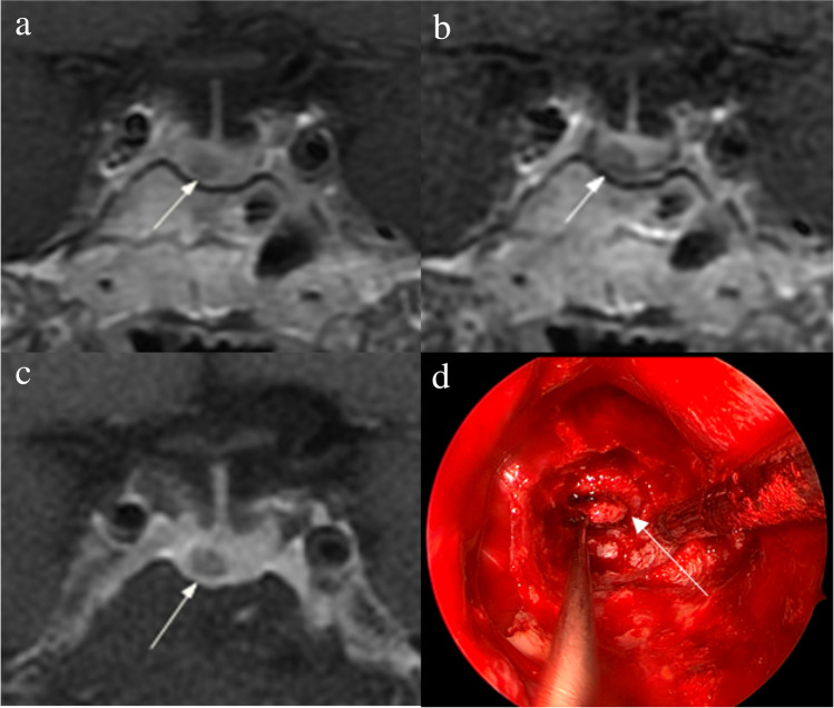
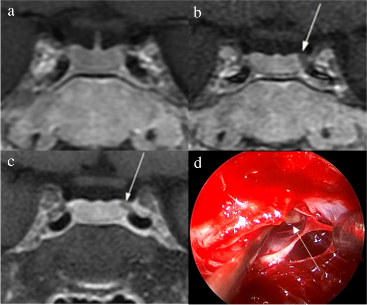
To assess the diagnostic performance of high-resolution contrast-enhanced MRI (hrMRI) with three-dimensional (3D) fast spin echo (FSE) sequence by comparison with conventional contrast-enhanced MRI (cMRI) and dynamic contrast-enhanced MRI (dMRI) with 2D FSE sequence for identifying pituitary microadenomas.

METHODS

This single-institutional retrospective study included 69 consecutive patients with Cushing's syndrome who underwent preoperative pituitary MRI, including cMRI, dMRI, and hrMRI, between January 2016 to December 2020. Reference standards were established by using all available imaging, clinical, surgical, and pathological resources. The diagnostic performance of cMRI, dMRI, and hrMRI for identifying pituitary microadenomas was independently evaluated by two experienced neuroradiologists. The area under the receiver operating characteristics curves (AUCs) were compared between protocols for each reader by using the DeLong test to assess the diagnostic performance for identifying pituitary microadenomas. The inter-observer agreement was assessed by using the κ analysis.

RESULTS

The diagnostic performance of hrMRI (AUC, 0.95-0.97) was higher than cMRI (AUC, 0.74-0.75; p ≤ .002) and dMRI (AUC, 0.59-0.68; p ≤ .001) for identifying pituitary microadenomas. The sensitivity and specificity of hrMRI were 90-93% and 100%, respectively. There were 78% (18/23) to 82% (14/17) of the patients, who were misdiagnosed on cMRI and dMRI and correctly diagnosed on hrMRI. The inter-observer agreement for identifying pituitary microadenomas was moderate on cMRI (κ = 0.50), moderate on dMRI (κ = 0.57), and almost perfect on hrMRI (κ = 0.91), respectively.

CONCLUSIONS

The hrMRI showed higher diagnostic performance than cMRI and dMRI for identifying pituitary microadenomas in patients with Cushing's syndrome.

KEY POINTS

• The diagnostic performance of hrMRI was higher than cMRI and dMRI for identifying pituitary microadenomas in Cushing's syndrome. • About 80% of patients, who were misdiagnosed on cMRI and dMRI, were correctly diagnosed on hrMRI. • The inter-observer agreement for identifying pituitary microadenomas was almost perfect on hrMRI.

摘要

目的

通过与常规对比增强磁共振成像(cMRI)和二维快速自旋回波(2D FSE)序列动态对比增强磁共振成像(dMRI)比较,评估三维(3D)快速自旋回波(FSE)序列高分辨率对比增强磁共振成像(hrMRI)对识别垂体微腺瘤的诊断性能。

方法

这项单机构回顾性研究纳入了 2016 年 1 月至 2020 年 12 月期间 69 例经术前垂体 MRI(包括 cMRI、dMRI 和 hrMRI)检查诊断为库欣综合征的连续患者。参考标准通过使用所有可用的影像学、临床、手术和病理资源建立。两名有经验的神经放射科医生独立评估 cMRI、dMRI 和 hrMRI 识别垂体微腺瘤的诊断性能。使用 DeLong 检验比较每个读者不同方案的受试者工作特征曲线下面积(AUC),以评估识别垂体微腺瘤的诊断性能。使用κ分析评估观察者间一致性。

结果

hrMRI(AUC:0.95-0.97)的诊断性能高于 cMRI(AUC:0.74-0.75;p≤0.002)和 dMRI(AUC:0.59-0.68;p≤0.001),用于识别垂体微腺瘤。hrMRI 的敏感性和特异性分别为 90-93%和 100%。在 cMRI 和 dMRI 误诊的 23 例患者中有 78%(18/23)至 82%(14/17)在 hrMRI 上得到正确诊断。cMRI 识别垂体微腺瘤的观察者间一致性为中度(κ=0.50),dMRI 为中度(κ=0.57),hrMRI 为极好(κ=0.91)。

结论

在库欣综合征患者中,hrMRI 对识别垂体微腺瘤的诊断性能优于 cMRI 和 dMRI。

https://cdn.ncbi.nlm.nih.gov/pmc/blobs/e9e9/10415427/bd5543eaa13a/330_2023_9585_Fig5_HTML.jpg
https://cdn.ncbi.nlm.nih.gov/pmc/blobs/e9e9/10415427/5c7eabc0e206/330_2023_9585_Fig1_HTML.jpg
https://cdn.ncbi.nlm.nih.gov/pmc/blobs/e9e9/10415427/b332a8ebd317/330_2023_9585_Fig2_HTML.jpg
https://cdn.ncbi.nlm.nih.gov/pmc/blobs/e9e9/10415427/9fd794466750/330_2023_9585_Fig3_HTML.jpg
https://cdn.ncbi.nlm.nih.gov/pmc/blobs/e9e9/10415427/fcd4451f6988/330_2023_9585_Fig4_HTML.jpg
https://cdn.ncbi.nlm.nih.gov/pmc/blobs/e9e9/10415427/bd5543eaa13a/330_2023_9585_Fig5_HTML.jpg

相似文献

1
High-resolution contrast-enhanced MRI with three-dimensional fast spin echo improved the diagnostic performance for identifying pituitary microadenomas in Cushing's syndrome.高分辨率对比增强磁共振成像结合三维快速自旋回波可提高库欣综合征中垂体微腺瘤的诊断性能。
Eur Radiol. 2023 Sep;33(9):5984-5992. doi: 10.1007/s00330-023-09585-1. Epub 2023 May 22.
2
Three-Dimensional Fast Spin Echo Pituitary MRI in Treatment-Naïve Cushing's Disease: Reduced Impact of Reader Experience and Increased Diagnostic Accuracy.治疗初发库欣病的三维快速自旋回波垂体 MRI:降低了读者经验的影响,提高了诊断准确性。
J Magn Reson Imaging. 2024 Jun;59(6):2115-2123. doi: 10.1002/jmri.28975. Epub 2023 Sep 1.
3
Comparative evaluation of conventional and dynamic magnetic resonance imaging of the pituitary gland for the diagnosis of Cushing's disease.垂体常规磁共振成像与动态磁共振成像对库欣病诊断的比较评估
Clin Endocrinol (Oxf). 1998 Sep;49(3):293-300. doi: 10.1046/j.1365-2265.1998.00541.x.
4
Comparison of MRI techniques for detecting microadenomas in Cushing's disease.比较磁共振成像技术在库欣病微腺瘤检测中的应用。
J Neurosurg. 2018 Apr;128(4):1051-1057. doi: 10.3171/2017.3.JNS163122. Epub 2017 Apr 28.
5
Potential utility of FLAIR in MRI-negative Cushing's disease.FLAIR 在 MRI 阴性库欣病中的潜在应用。
J Neurosurg. 2018 Sep;129(3):620-628. doi: 10.3171/2017.4.JNS17234. Epub 2017 Oct 13.
6
High-dose dexamethasone suppression test is inferior to pituitary dynamic enhanced MRI in the differential diagnosis of ACTH-dependent Cushing's syndrome.大剂量地塞米松抑制试验在鉴别诊断 ACTH 依赖性库欣综合征方面不如垂体动态增强 MRI。
Endocrine. 2022 Feb;75(2):516-524. doi: 10.1007/s12020-021-02891-y. Epub 2021 Oct 4.
7
Volume interpolated 3D-spoiled gradient echo sequence is better than dynamic contrast spin echo sequence for MRI detection of corticotropin secreting pituitary microadenomas.容积内插 3D 扰相梯度回波序列优于动态对比自旋回波序列,用于 MRI 检测促肾上腺皮质激素分泌垂体微腺瘤。
Clin Endocrinol (Oxf). 2013 Jun;78(6):825-30. doi: 10.1111/cen.12069. Epub 2013 Apr 6.
8
Cavernous and inferior petrosal sinus sampling and dynamic magnetic resonance imaging in the preoperative evaluation of Cushing's disease.海绵窦和岩下窦取样及动态磁共振成像在库欣病术前评估中的应用。
J Neurooncol. 2014 Feb;116(3):593-600. doi: 10.1007/s11060-013-1342-9. Epub 2014 Jan 8.
9
Pituitary tumour localization in patients with Cushing's disease by magnetic resonance imaging. Is there a place for petrosal sinus sampling?库欣病患者垂体瘤的磁共振成像定位。岩下窦采血还有用吗?
Clin Endocrinol (Oxf). 1994 Jan;40(1):87-92. doi: 10.1111/j.1365-2265.1994.tb02448.x.
10
Emerging diagnostic methods and imaging modalities in cushing's syndrome.库欣综合征的新兴诊断方法和影像学手段。
Front Endocrinol (Lausanne). 2023 Jul 25;14:1230447. doi: 10.3389/fendo.2023.1230447. eCollection 2023.

引用本文的文献

1
Improved noninvasive diagnostic evaluations in treatment-naïve adrenocorticotropic hormone (ACTH)-dependent Cushing's syndrome.初治促肾上腺皮质激素(ACTH)依赖性库欣综合征的无创诊断评估改善
BMC Med Imaging. 2025 Jul 1;25(1):252. doi: 10.1186/s12880-025-01786-y.
2
Evaluation of high-resolution pituitary dynamic contrast-enhanced MRI using deep learning-based compressed sensing and super-resolution reconstruction.基于深度学习的压缩感知和超分辨率重建对高分辨率垂体动态对比增强磁共振成像的评估
Eur Radiol. 2025 Apr 13. doi: 10.1007/s00330-025-11574-5.
3
A semantic segmentation model for automatic precise identification of pituitary microadenomas with preoperative MRI.

本文引用的文献

1
Thin-Slice Pituitary MRI with Deep Learning-based Reconstruction: Diagnostic Performance in a Postoperative Setting.基于深度学习重建的垂体薄层 MRI:在术后环境中的诊断性能。
Radiology. 2021 Jan;298(1):114-122. doi: 10.1148/radiol.2020200723. Epub 2020 Nov 3.
2
Compressed SENSE accelerated 3D T1w black blood turbo spin echo versus 2D T1w turbo spin echo sequence in pituitary magnetic resonance imaging.压缩感知加速 3D T1w 黑血 turbo 自旋回波与 2D T1w turbo 自旋回波序列在垂体磁共振成像中的比较。
Eur J Radiol. 2019 Nov;120:108667. doi: 10.1016/j.ejrad.2019.108667. Epub 2019 Sep 14.
3
Outcomes of three-Tesla magnetic resonance imaging for the identification of pituitary adenoma in patients with Cushing's disease.
一种用于通过术前磁共振成像自动精确识别垂体微腺瘤的语义分割模型。
Neuroradiology. 2025 Apr;67(4):1061-1070. doi: 10.1007/s00234-025-03599-w. Epub 2025 Apr 4.
3特斯拉磁共振成像用于库欣病患者垂体腺瘤识别的结果
Endocr J. 2019 Mar 28;66(3):259-264. doi: 10.1507/endocrj.EJ18-0458. Epub 2019 Feb 14.
4
Comparison of MRI techniques for detecting microadenomas in Cushing's disease.比较磁共振成像技术在库欣病微腺瘤检测中的应用。
J Neurosurg. 2018 Apr;128(4):1051-1057. doi: 10.3171/2017.3.JNS163122. Epub 2017 Apr 28.
5
Diagnosis and Differential Diagnosis of Cushing's Syndrome.库欣综合征的诊断与鉴别诊断
N Engl J Med. 2017 Apr 13;376(15):1451-1459. doi: 10.1056/NEJMra1505550.
6
Pituitary magnetic resonance imaging in Cushing's disease.库欣病的垂体磁共振成像
Endocrine. 2017 Mar;55(3):691-696. doi: 10.1007/s12020-016-1038-y. Epub 2016 Jul 19.
7
Cushing's disease: pathobiology, diagnosis, and management.库欣病:病理生物学、诊断与治疗。
J Neurosurg. 2017 Feb;126(2):404-417. doi: 10.3171/2016.1.JNS152119. Epub 2016 Apr 22.
8
Treatment of Cushing's Syndrome: An Endocrine Society Clinical Practice Guideline.库欣综合征的治疗:美国内分泌学会临床实践指南
J Clin Endocrinol Metab. 2015 Aug;100(8):2807-31. doi: 10.1210/jc.2015-1818. Epub 2015 Jul 29.
9
Cushing's syndrome.库欣综合征。
Lancet. 2015 Aug 29;386(9996):913-27. doi: 10.1016/S0140-6736(14)61375-1. Epub 2015 May 21.
10
Three-Tesla imaging of the pituitary and parasellar region: T1-weighted 3-dimensional fast spin echo cube outperforms conventional 2-dimensional magnetic resonance imaging.垂体及鞍旁区域的3特斯拉成像:T1加权三维快速自旋回波立方序列优于传统二维磁共振成像。
J Comput Assist Tomogr. 2015 May-Jun;39(3):329-33. doi: 10.1097/RCT.0000000000000214.