Vita-Salute San Raffaele University, Via Olgettina 60, Milan, Italy.
Department of Ophthalmology, IRCCS San Raffaele Scientific Institute, Milan, Italy.
Sci Rep. 2023 May 22;13(1):8237. doi: 10.1038/s41598-023-35339-6.
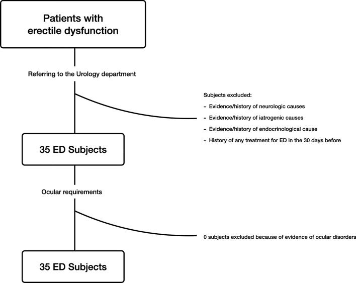
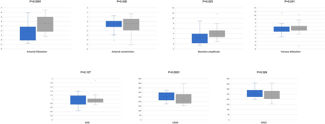
The aim of this study was to study the retinal vessels in patients affected by vasculogenic erectile dysfunction (ED), using dynamic vessel analyzer (DVA). Patients with vasculogenic ED and control subjects were prospectively enrolled to undergo a complete urological and ophthalmologic evaluation, including DVA and structural optical coherence tomography (OCT). The main outcome measures were: (1) arterial dilation; (2) arterial constriction; (3) reaction amplitude (the difference between arterial dilation and constriction); and, (4) venous dilation. Thirty-five patients with ED and 30 male controls were included in the analysis. Mean ± SD age was 52.0 ± 10.8 years in the ED group and 48.1 ± 16.3 years in the control group (p = 0.317). In the dynamic analysis, the arterial dilation was lower in the ED group (1.88 ± 1.50%), as compared with the control group (3.70 ± 1.56%, p < 0.0001). Neither arterial constriction nor venous dilation differed between groups. The reaction amplitude was decreased in ED patients (2.40 ± 2.02%, p = 0.023), compared to controls (4.25 ± 2.20%). In the Pearson correlation analysis, the ED severity, was directly correlated with both reaction amplitude (R = .701, p = 0.004) and arterial dilation (R = .529, p = 0.042). In conclusion, subjects with vasculogenic ED are featured by a significant dysfunction of the retinal neurovascular coupling, which is inversely correlated with ED severity.
本研究旨在使用动态血管分析仪(DVA)研究血管性勃起功能障碍(ED)患者的视网膜血管。前瞻性纳入血管性 ED 患者和对照组患者,进行全面的泌尿科和眼科评估,包括 DVA 和结构光相干断层扫描(OCT)。主要观察指标为:(1)动脉扩张;(2)动脉收缩;(3)反应幅度(动脉扩张和收缩之间的差异);和(4)静脉扩张。分析纳入 35 例 ED 患者和 30 名男性对照组。ED 组的平均年龄为 52.0±10.8 岁,对照组为 48.1±16.3 岁(p=0.317)。在动态分析中,ED 组的动脉扩张较低(1.88±1.50%),与对照组(3.70±1.56%)相比,差异有统计学意义(p<0.0001)。两组间动脉收缩和静脉扩张无差异。ED 患者的反应幅度降低(2.40±2.02%),与对照组(4.25±2.20%)相比,差异有统计学意义(p=0.023)。Pearson 相关分析显示,ED 严重程度与反应幅度(R=0.701,p=0.004)和动脉扩张(R=0.529,p=0.042)呈正相关。总之,血管性 ED 患者的视网膜神经血管耦合功能明显受损,且与 ED 严重程度呈负相关。