Bansal Akash, Kumar Sushil, Rai Neha, Kumari Shilpi, Kumar Visesh, Kumar Ajeet, Chandra Nimai Chand
Patna, 801507 India Department of Biochemistry, All India Institute of Medical Sciences.
Greater Noida, Gautam Budh Nagar, Uttar Pradesh 201301 India Department of Biochemistry, School of Basic Applied Sciences, Galgotias University.
Indian J Clin Biochem. 2023 Jul;38(3):374-384. doi: 10.1007/s12291-023-01121-8. Epub 2023 Mar 9.
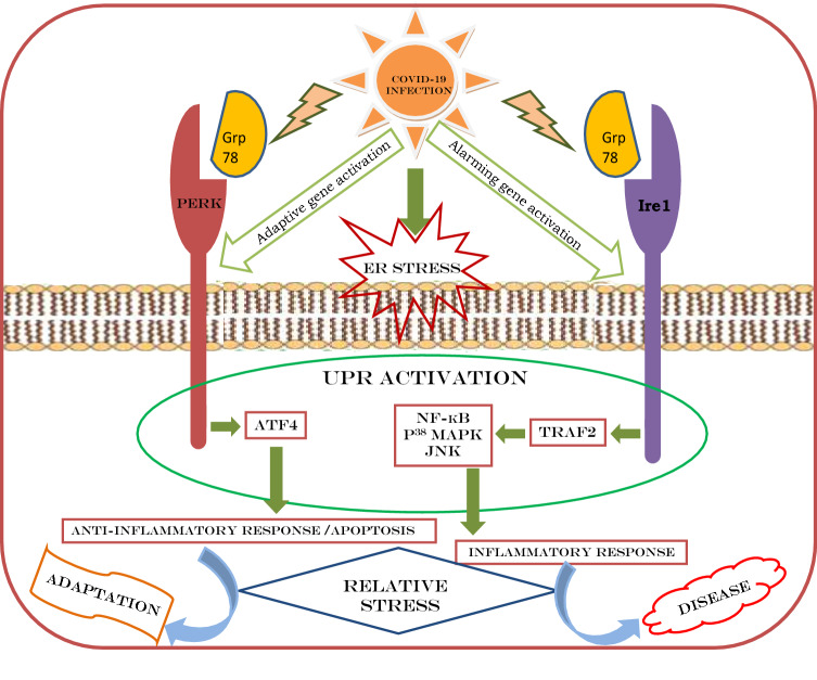
The endoplasmic reticulum (ER) is the site for protein synthesis, its folding and secretion. An intricate set of signalling pathways, called UPR pathways, have been evolved by ER in mammalian cells, to allow the cell to respond the presence of misfolded proteins within the ER. Breaching of these signalling systems by disease oriented accumulation of unfolded proteins may develop cellular stress. The aim of this study is to explore whether COVID-19 infection is responsible for developing this kind of endoplasmic reticulum related stress (ER-stress). ER-stress was evaluated by checking the expression of ER-stress markers e.g. PERK (adapting) and TRAF2 (alarming). ER-stress was correlated to several blood parameters viz. IgG, pro- and anti-inflammatory cytokines, leukocytes, lymphocytes, RBC, haemoglobin and PaO/FiO ratio (ratio of arterial oxygen partial pressure to fractional inspired oxygen) in COVID-19 affected subjects. COVID-19 infection was found to be a state of protein homeostasis (proteostasis) collapse. Changes in IgG levels showed very poor immune response by the infected subjects. At the initial phase of the disease, pro-inflammatory cytokine levels were high and anti-inflammatory cytokines levels were low; though they were partly compromised at later phase of the disease. Total leukocyte concentration increased over the period of time; while percentage of lymphocytes were dropped. No significant changes were observed in cases of RBC counts and haemoglobin (Hb) levels. Both RBC and Hb were maintained at their normal range. In mildly stressed group, PaO/FiO ratio (oxygenation status) was in the higher side of normal range; whereas in other two groups the ratio was in respiratory distress syndrome mode. Virus could induce mild to severe ER-stress, which could be the cause of cellular death and systemic dysfunction introducing fatal consequences.
Schematic representation of SARS-CoV-2 infection and related consequences.
内质网(ER)是蛋白质合成、折叠和分泌的场所。在哺乳动物细胞中,内质网进化出了一套复杂的信号通路,称为未折叠蛋白反应(UPR)通路,以使细胞能够应对内质网中错误折叠蛋白的存在。疾病导向的未折叠蛋白积累破坏这些信号系统可能会导致细胞应激。本研究的目的是探讨新型冠状病毒肺炎(COVID-19)感染是否会导致这种内质网相关应激(ER应激)。通过检测ER应激标志物如PERK(适应)和TRAF2(警报)的表达来评估ER应激。在COVID-19感染患者中,ER应激与多种血液参数相关,即免疫球蛋白G(IgG)、促炎和抗炎细胞因子、白细胞、淋巴细胞、红细胞、血红蛋白以及动脉血氧分压与吸入氧分数之比(PaO/FiO比值)。发现COVID-19感染是一种蛋白质稳态(蛋白平衡)崩溃的状态。IgG水平的变化表明感染患者的免疫反应非常差。在疾病的初始阶段,促炎细胞因子水平高,抗炎细胞因子水平低;尽管在疾病后期它们部分受损。随着时间的推移,白细胞总数增加;而淋巴细胞百分比下降。红细胞计数和血红蛋白(Hb)水平未观察到显著变化。红细胞和血红蛋白均维持在正常范围内。在轻度应激组中,PaO/FiO比值(氧合状态)处于正常范围的较高水平;而在其他两组中,该比值处于呼吸窘迫综合征模式。病毒可诱导轻度至重度的ER应激,这可能是细胞死亡和全身功能障碍的原因,从而导致致命后果。
严重急性呼吸综合征冠状病毒2(SARS-CoV-2)感染及相关后果的示意图。