Department of Gastroenterological Surgery, Hirosaki University, Graduate School of Medicine, Hirosaki, Aomori, Japan.
Department of Anatomical Science, Hirosaki University, Graduate School of Medicine, Hirosaki, Aomori, Japan.
PLoS One. 2023 May 25;18(5):e0286316. doi: 10.1371/journal.pone.0286316. eCollection 2023.
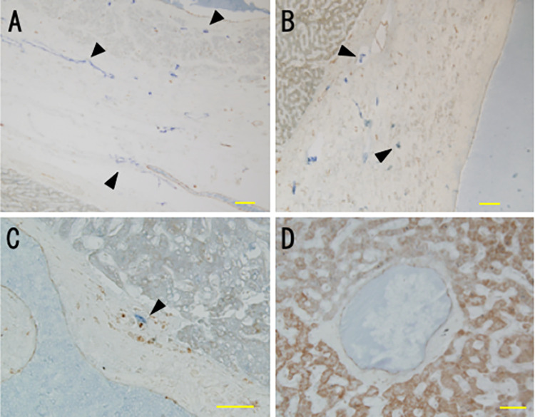
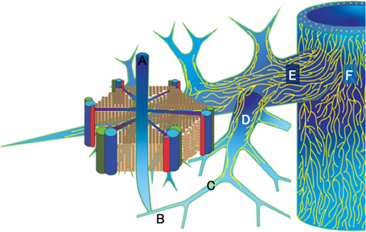
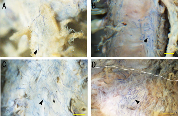
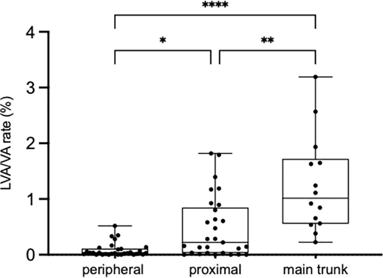
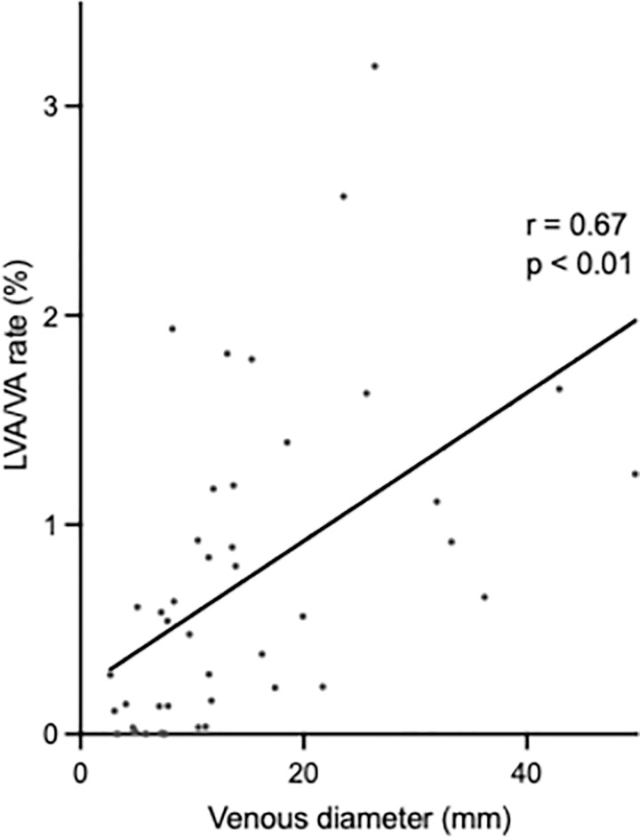
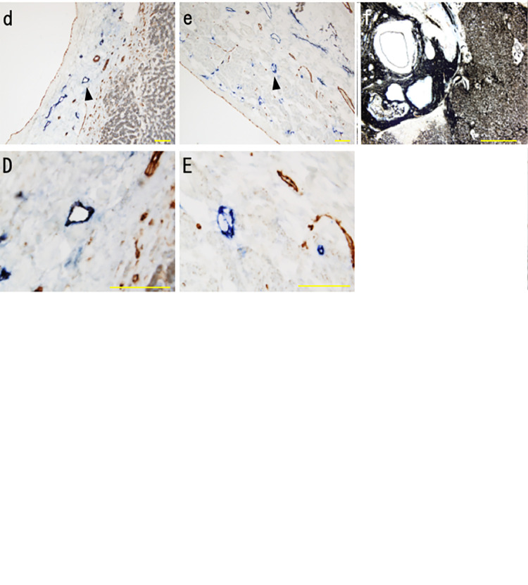
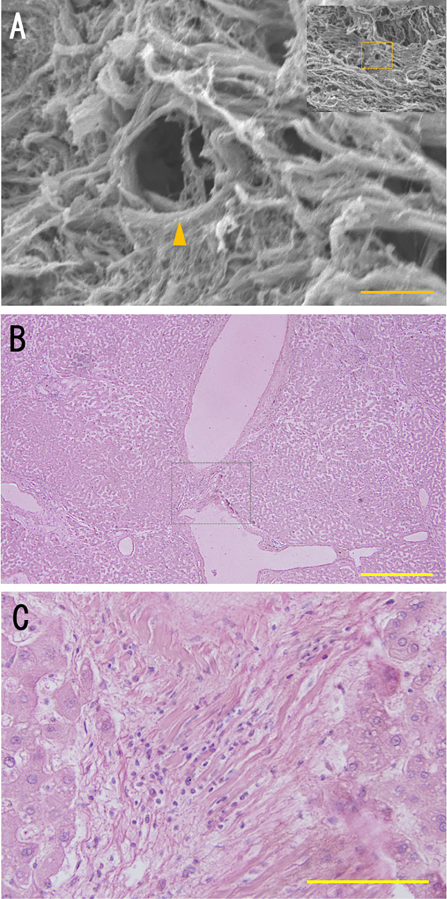
Lymphatic fluid drains from the liver via the periportal lymphatic, hepatic venous lymphatic, and superficial lymphatic systems. We performed a postmortem study to clarify the three-dimensional structure and flow dynamics of the human hepatic venous lymphatic system, as it still remains unclear. Livers were excised whole from three human cadavers, injected with India ink, and sliced into 1-cm sections from which veins were harvested. The distribution of lymphatic vessels was observed in 5 μm sections immunostained for lymphatic and vascular markers (podoplanin and CD31, respectively) using light microscopy. Continuity and density of lymphatic vessel distribution were assessed in en-face whole-mount preparations of veins using stereomicroscopy. The structure of the external hepatic vein wall was assessed with scanning electron microscopy (SEM). The lymphatic dynamics study suggested that lymphatic fluid flows through an extravascular pathway around the central and sublobular veins. A lymphatic vessel network originates in the wall of sublobular veins, with a diameter greater than 110 μm, and the peripheral portions of hepatic veins and continues to the inferior vena cava. The density distribution of lymphatic vessels is smallest in the peripheral portion of the hepatic vein (0.03%) and increases to the proximal portion (0.22%, p = 0.012) and the main trunk (1.01%, p < 0.001), correlating positively with increasing hepatic vein diameter (Rs = 0.67, p < 0.001). We revealed the three-dimensional structure of the human hepatic venous lymphatic system. The results could improve the understanding of lymphatic physiology and liver pathology.
淋巴液通过门脉周围淋巴管、肝静脉淋巴管和浅表淋巴管系统从肝脏排出。我们进行了一项尸检研究,以阐明人类肝静脉淋巴管系统的三维结构和流动动力学,因为其仍然不清楚。从三个人体尸检中取出整个肝脏,注入印度墨水,并切成 1 厘米的切片,从这些切片中取出静脉。使用淋巴和血管标志物(分别为 podoplanin 和 CD31)对 5 μm 切片进行免疫染色,观察淋巴管的分布。使用体视显微镜观察静脉的全面立体准备,评估淋巴管分布的连续性和密度。使用扫描电子显微镜(SEM)评估肝外静脉壁的结构。淋巴动力学研究表明,淋巴液通过围绕中央和小叶静脉的血管外途径流动。淋巴管网络起源于小叶静脉壁,直径大于 110 μm,并且存在于肝静脉和下腔静脉的周围部分。淋巴管的密度分布在肝静脉的周围部分最小(0.03%),并增加到近端部分(0.22%,p = 0.012)和主干(1.01%,p < 0.001),与肝静脉直径的增加呈正相关(Rs = 0.67,p < 0.001)。我们揭示了人类肝静脉淋巴管系统的三维结构。这些结果可以提高对淋巴生理学和肝脏病理学的理解。