Reddy-Vari Hymavathi, Kim Yerin, Rajput Charu, Sajjan Umadevi S
Department of Microbiology Immunology and Inflammation, Lewis Katz Medical School, Temple University, Philadelphia, PA, USA.
Department of Thoracic Surgery and Medicine, Lewis Katz Medical School, Temple University, Philadelphia, PA, USA.
ERJ Open Res. 2023 May 22;9(3). doi: 10.1183/23120541.00694-2022. eCollection 2023 May.
Airway epithelial cells from patients with COPD show suboptimal innate immune responses to nontypeable (NTHi) and Toll-like receptor (TLR)2 ligands despite expressing TLR2 similar to normal airway epithelial cells, but the underlying mechanisms are poorly understood.
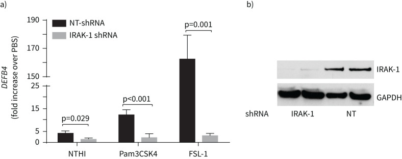
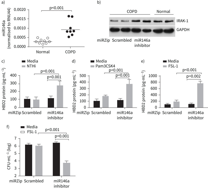
Normal or COPD mucociliary-differentiated airway epithelial cells were treated with TLR2 agonists or infected with NTHi and expression of β-defensin (HBD)2 was examined. Interleukin-1 receptor-associated kinase (IRAK)-1 and microRNA (miR)146a were genetically inhibited in normal and COPD airway epithelial cell cultures, respectively, and HBD2 responses to TLR2 ligands were determined. IRAK-1 expression in lung sections was determined by immunofluorescence microscopy.
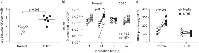
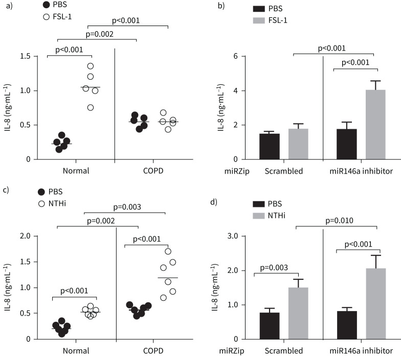
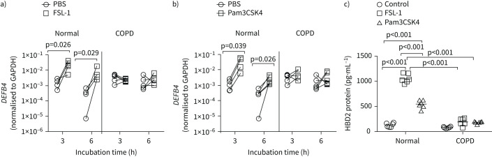
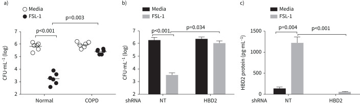
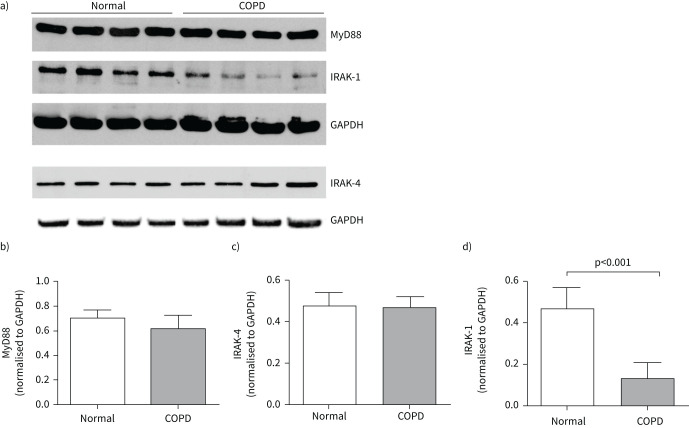
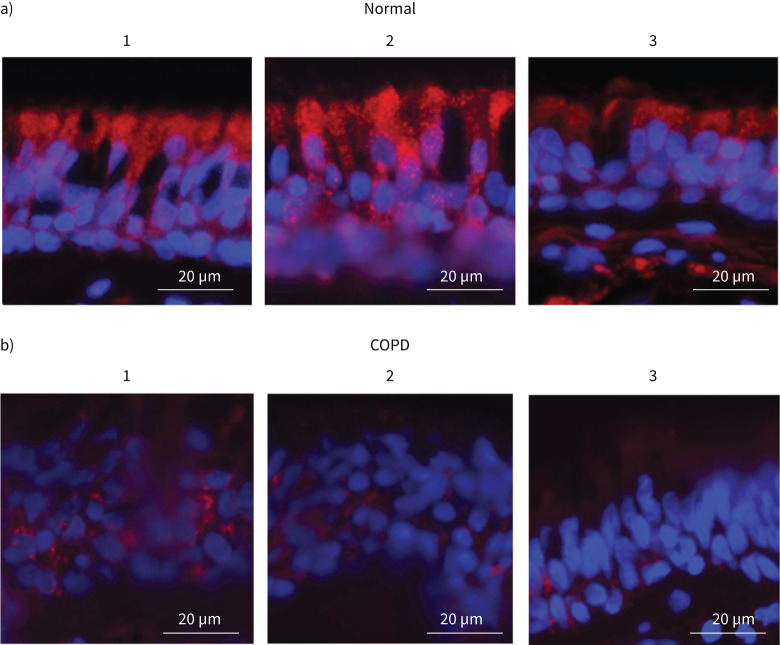
Compared to normal, COPD airway epithelial cell cultures showed impaired expression of HBD2 in response to TLR2 agonists or NTHi infection. Apical secretions from TLR2 agonist-treated normal, but not COPD, airway epithelial cells efficiently killed NTHi. Knockdown of HBD2 significantly reduced NTHi killing by apical secretions of normal airway epithelial cells. Compared to normal, COPD cells showed significantly reduced expression of IRAK-1 and this was associated with increased expression of miR146a. Inhibition of miR146a increased the expression of IRAK-1, improved the expression of HBD2 in response to TLR2 agonists in COPD cells and enhanced the killing of bacteria by apical secretions obtained from TLR2 agonist-treated COPD cells. Bronchial epithelium of COPD patients showed reduced expression of IRAK-1.
These results suggest that reduced levels of IRAK-1 due to increased expression of miR146a may contribute to impaired expression of TLR2-induced HBD2 in COPD airway epithelial cells.
慢性阻塞性肺疾病(COPD)患者的气道上皮细胞对不可分型流感嗜血杆菌(NTHi)和Toll样受体(TLR)2配体的先天性免疫反应欠佳,尽管其TLR2表达与正常气道上皮细胞相似,但潜在机制尚不清楚。
用TLR2激动剂处理正常或COPD黏液纤毛分化的气道上皮细胞,或用NTHi感染,检测β-防御素(HBD)2的表达。分别在正常和COPD气道上皮细胞培养物中对白细胞介素-1受体相关激酶(IRAK)-1和微小RNA(miR)146a进行基因抑制,测定HBD2对TLR2配体的反应。通过免疫荧光显微镜测定肺切片中IRAK-1的表达。
与正常气道上皮细胞培养物相比,COPD气道上皮细胞培养物对TLR2激动剂或NTHi感染的反应中,HBD2表达受损。经TLR2激动剂处理的正常气道上皮细胞而非COPD气道上皮细胞的顶端分泌物能有效杀灭NTHi。敲低HBD2可显著降低正常气道上皮细胞顶端分泌物对NTHi的杀灭作用。与正常细胞相比,COPD细胞中IRAK-1的表达显著降低,这与miR146a表达增加有关。抑制miR146a可增加IRAK-1的表达,改善COPD细胞中TLR2激动剂刺激下HBD2的表达,并增强经TLR2激动剂处理的COPD细胞顶端分泌物对细菌的杀灭作用。COPD患者的支气管上皮中IRAK-1表达降低。
这些结果表明,miR146a表达增加导致的IRAK-1水平降低可能是COPD气道上皮细胞中TLR2诱导的HBD2表达受损的原因。