• 文献检索
  • 文档翻译
  • 深度研究
  • 学术资讯
  • Suppr Zotero 插件Zotero 插件
  • 邀请有礼
  • 套餐&价格
  • 历史记录
应用&插件
Suppr Zotero 插件Zotero 插件浏览器插件Mac 客户端Windows 客户端微信小程序
定价
高级版会员购买积分包购买API积分包
服务
文献检索文档翻译深度研究API 文档MCP 服务
关于我们
关于 Suppr公司介绍联系我们用户协议隐私条款
关注我们

Suppr 超能文献

核心技术专利:CN118964589B侵权必究
粤ICP备2023148730 号-1Suppr @ 2026

文献检索

告别复杂PubMed语法,用中文像聊天一样搜索,搜遍4000万医学文献。AI智能推荐,让科研检索更轻松。

立即免费搜索

文件翻译

保留排版,准确专业,支持PDF/Word/PPT等文件格式,支持 12+语言互译。

免费翻译文档

深度研究

AI帮你快速写综述,25分钟生成高质量综述,智能提取关键信息,辅助科研写作。

立即免费体验

催乳素受体通过与磷酸肌醇-4,5-二磷酸共构形成来支架连接激酶 2。

The prolactin receptor scaffolds Janus kinase 2 via co-structure formation with phosphoinositide-4,5-bisphosphate.

机构信息

Structural Biophysics, Section for Neutron and X-ray Science, Niels Bohr Institute, University of Copenhagen, Copenhagen, Denmark.

Facultad de Ingenieria Arquitectura y Diseño, Universidad San Sebastian, Santiago, Chile.

出版信息

Elife. 2023 May 26;12:e84645. doi: 10.7554/eLife.84645.

DOI:10.7554/eLife.84645
PMID:37232489
原文链接:https://pmc.ncbi.nlm.nih.gov/articles/PMC10260020/
Abstract

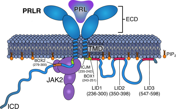
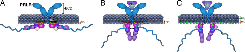
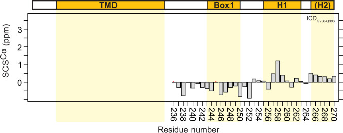
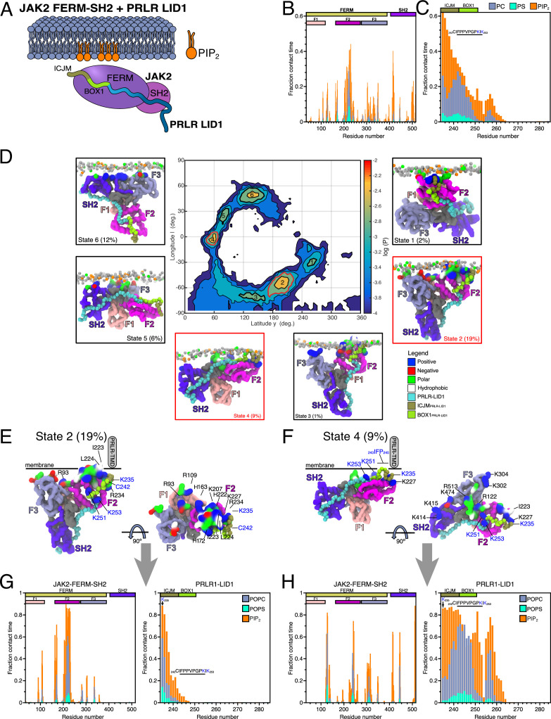
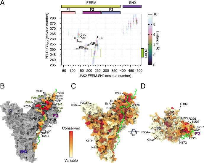
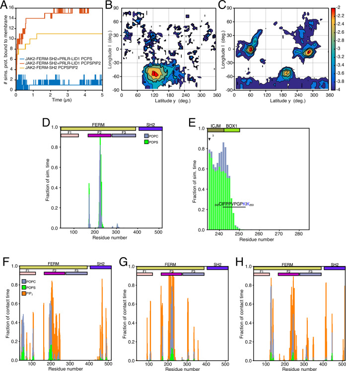
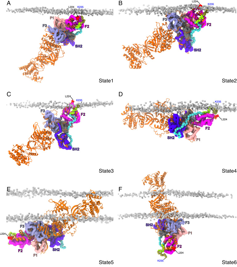
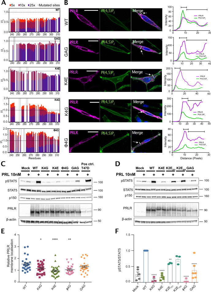
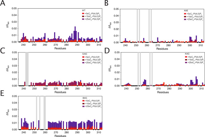
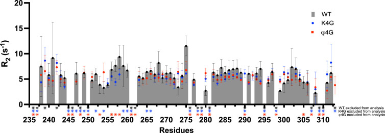
Class 1 cytokine receptors transmit signals through the membrane by a single transmembrane helix to an intrinsically disordered cytoplasmic domain that lacks kinase activity. While specific binding to phosphoinositides has been reported for the prolactin receptor (PRLR), the role of lipids in PRLR signaling is unclear. Using an integrative approach combining nuclear magnetic resonance spectroscopy, cellular signaling experiments, computational modeling, and simulation, we demonstrate co-structure formation of the disordered intracellular domain of the human PRLR, the membrane constituent phosphoinositide-4,5-bisphosphate (PI(4,5)P) and the FERM-SH2 domain of the Janus kinase 2 (JAK2). We find that the complex leads to accumulation of PI(4,5)P at the transmembrane helix interface and that the mutation of residues identified to interact specifically with PI(4,5)P negatively affects PRLR-mediated activation of signal transducer and activator of transcription 5 (STAT5). Facilitated by co-structure formation, the membrane-proximal disordered region arranges into an extended structure. We suggest that the co-structure formed between PRLR, JAK2, and PI(4,5)P locks the juxtamembrane disordered domain of the PRLR in an extended structure, enabling signal relay from the extracellular to the intracellular domain upon ligand binding. We find that the co-structure exists in different states which we speculate could be relevant for turning signaling on and off. Similar co-structures may be relevant for other non-receptor tyrosine kinases and their receptors.

摘要

I 类细胞因子受体通过单一跨膜螺旋将信号传递到无激酶活性的内在无序胞质域。虽然已报道催乳素受体 (PRLR) 与磷酸肌醇结合,但脂质在 PRLR 信号中的作用尚不清楚。我们采用综合方法,结合核磁共振波谱、细胞信号实验、计算建模和模拟,证明了人类 PRLR 的无序胞内域、膜成分磷酸肌醇-4,5-二磷酸 (PI(4,5)P) 和 Janus 激酶 2 (JAK2) 的 FERM-SH2 结构域的共结构形成。我们发现该复合物导致 PI(4,5)P 在跨膜螺旋界面处积累,并且与 PI(4,5)P 特异性相互作用的残基的突变会负影响 PRLR 介导的信号转导和转录激活物 5 (STAT5) 的激活。共结构形成促进了膜近端无序区域的延伸结构排列。我们认为,PRLR、JAK2 和 PI(4,5)P 之间形成的共结构将 PRLR 的跨膜近侧无序域锁定在延伸结构中,从而在配体结合后,能够将信号从细胞外传递到细胞内结构域。我们发现该共结构存在于不同的状态,我们推测这可能与信号的开启和关闭有关。类似的共结构可能与其他非受体酪氨酸激酶及其受体有关。

https://cdn.ncbi.nlm.nih.gov/pmc/blobs/5237/10260020/24043189e887/elife-84645-fig6.jpg
https://cdn.ncbi.nlm.nih.gov/pmc/blobs/5237/10260020/fcac3f7fed7f/elife-84645-fig1.jpg
https://cdn.ncbi.nlm.nih.gov/pmc/blobs/5237/10260020/501791648f5d/elife-84645-fig2.jpg
https://cdn.ncbi.nlm.nih.gov/pmc/blobs/5237/10260020/8e4c2954b463/elife-84645-fig2-figsupp1.jpg
https://cdn.ncbi.nlm.nih.gov/pmc/blobs/5237/10260020/3701552edde1/elife-84645-fig2-figsupp2.jpg
https://cdn.ncbi.nlm.nih.gov/pmc/blobs/5237/10260020/4d68d682554f/elife-84645-fig3.jpg
https://cdn.ncbi.nlm.nih.gov/pmc/blobs/5237/10260020/c0a624620a9d/elife-84645-fig3-figsupp1.jpg
https://cdn.ncbi.nlm.nih.gov/pmc/blobs/5237/10260020/101b282e6e8f/elife-84645-fig3-figsupp2.jpg
https://cdn.ncbi.nlm.nih.gov/pmc/blobs/5237/10260020/e470cfbdd917/elife-84645-fig4.jpg
https://cdn.ncbi.nlm.nih.gov/pmc/blobs/5237/10260020/716307dc993a/elife-84645-fig4-figsupp1.jpg
https://cdn.ncbi.nlm.nih.gov/pmc/blobs/5237/10260020/3aece91c77aa/elife-84645-fig4-figsupp2.jpg
https://cdn.ncbi.nlm.nih.gov/pmc/blobs/5237/10260020/074465dd48c7/elife-84645-fig4-figsupp3.jpg
https://cdn.ncbi.nlm.nih.gov/pmc/blobs/5237/10260020/00a7ae39d52f/elife-84645-fig5.jpg
https://cdn.ncbi.nlm.nih.gov/pmc/blobs/5237/10260020/449f04373e97/elife-84645-fig5-figsupp1.jpg
https://cdn.ncbi.nlm.nih.gov/pmc/blobs/5237/10260020/caed1faaed64/elife-84645-fig5-figsupp2.jpg
https://cdn.ncbi.nlm.nih.gov/pmc/blobs/5237/10260020/24043189e887/elife-84645-fig6.jpg

相似文献

1
The prolactin receptor scaffolds Janus kinase 2 via co-structure formation with phosphoinositide-4,5-bisphosphate.催乳素受体通过与磷酸肌醇-4,5-二磷酸共构形成来支架连接激酶 2。
Elife. 2023 May 26;12:e84645. doi: 10.7554/eLife.84645.
2
SOCS-1, -2, -3: selective targets and functions downstream of the prolactin receptor.细胞因子信号转导抑制因子-1、-2、-3:催乳素受体下游的选择性靶点及功能
Mol Cell Endocrinol. 1999 Dec 20;158(1-2):45-54. doi: 10.1016/s0303-7207(99)00180-x.
3
A non-catalytic function of the Src family tyrosine kinases controls prolactin-induced Jak2 signaling.Src 家族酪氨酸激酶的非催化功能控制催乳素诱导的 Jak2 信号转导。
Cell Signal. 2010 Mar;22(3):415-26. doi: 10.1016/j.cellsig.2009.10.013.
4
Growth hormone signaling in human T47D breast cancer cells: potential role for a growth hormone receptor-prolactin receptor complex.人T47D乳腺癌细胞中的生长激素信号传导:生长激素受体-催乳素受体复合物的潜在作用
Mol Endocrinol. 2011 Apr;25(4):597-610. doi: 10.1210/me.2010-0255. Epub 2011 Feb 10.
5
Prolactin receptor regulates Stat5 tyrosine phosphorylation and nuclear translocation by two separate pathways.催乳素受体通过两条独立的途径调节Stat5酪氨酸磷酸化和核转位。
J Biol Chem. 1998 Mar 27;273(13):7709-16. doi: 10.1074/jbc.273.13.7709.
6
Nectin-4 co-stimulates the prolactin receptor by interacting with SOCS1 and inhibiting its activity on the JAK2-STAT5a signaling pathway.NECTIN-4通过与SOCS1相互作用并抑制其对JAK2-STAT5a信号通路的活性来共刺激催乳素受体。
J Biol Chem. 2017 Apr 28;292(17):6895-6909. doi: 10.1074/jbc.M116.769091. Epub 2017 Mar 3.
7
Two wrongs can make a right: dimers of prolactin and growth hormone receptor antagonists behave as agonists.两个错误可为正确:催乳素二聚体与生长激素受体拮抗剂表现为激动剂。
Mol Endocrinol. 2006 Mar;20(3):661-74. doi: 10.1210/me.2005-0360. Epub 2005 Nov 3.
8
Diphenylbutylpiperidine Antipsychotic Drugs Inhibit Prolactin Receptor Signaling to Reduce Growth of Pancreatic Ductal Adenocarcinoma in Mice.二苯丁基哌啶类抗精神病药物通过抑制催乳素受体信号通路抑制胰腺导管腺癌在小鼠中的生长。
Gastroenterology. 2020 Apr;158(5):1433-1449.e27. doi: 10.1053/j.gastro.2019.11.279. Epub 2019 Nov 29.
9
The role of prolactin receptor in GH signaling in breast cancer cells.催乳素受体在乳腺癌细胞生长激素信号传导中的作用。
Mol Endocrinol. 2013 Feb;27(2):266-79. doi: 10.1210/me.2012-1297. Epub 2012 Nov 28.
10
Cytokine-inducible SH2-containing protein suppresses PRL signaling by binding the PRL receptor.细胞因子诱导含SH2结构域蛋白通过结合催乳素受体抑制催乳素信号传导。
Endocrinology. 2001 Dec;142(12):5286-93. doi: 10.1210/endo.142.12.8549.

引用本文的文献

1
Interactions Between Prolactin, Intracellular Signaling, and Possible Implications in the Contractility and Pathophysiology of Asthma.催乳素、细胞内信号传导之间的相互作用及其对哮喘收缩性和病理生理学的潜在影响
Int J Mol Sci. 2025 Jul 29;26(15):7332. doi: 10.3390/ijms26157332.
2
NEK kinases in cell cycle regulation, DNA damage response, and cancer progression.NEK激酶在细胞周期调控、DNA损伤反应及癌症进展中的作用
Tissue Cell. 2025 Jun;94:102811. doi: 10.1016/j.tice.2025.102811. Epub 2025 Feb 28.
3
Proline / Conformational Selection Controls 14-3-3 Binding.

本文引用的文献

1
Crosstalk between regulatory elements in disordered TRPV4 N-terminus modulates lipid-dependent channel activity.无规则 TRPV4 N 端调控元件间的串扰调节脂质依赖型通道活性。
Nat Commun. 2023 Jul 13;14(1):4165. doi: 10.1038/s41467-023-39808-4.
2
Tyrosine kinases compete for growth hormone receptor binding and regulate receptor mobility and degradation.酪氨酸激酶竞争生长激素受体结合并调节受体的流动性和降解。
Cell Rep. 2023 May 30;42(5):112490. doi: 10.1016/j.celrep.2023.112490. Epub 2023 May 9.
3
Different Forms of Disorder in NMDA-Sensitive Glutamate Receptor Cytoplasmic Domains Are Associated with Differences in Condensate Formation.
脯氨酸/构象选择控制14-3-3蛋白的结合。
J Am Chem Soc. 2025 Feb 19;147(7):5714-5724. doi: 10.1021/jacs.4c13462. Epub 2025 Feb 5.
4
The Influence of Phosphoinositide Lipids in the Molecular Biology of Membrane Proteins: Recent Insights from Simulations.磷酸肌醇脂质在膜蛋白分子生物学中的影响:来自模拟的最新见解
J Mol Biol. 2025 Feb 15;437(4):168937. doi: 10.1016/j.jmb.2025.168937. Epub 2025 Jan 9.
5
Charge-based immunoreceptor signalling in health and disease.健康与疾病中基于电荷的免疫受体信号传导。
Nat Rev Immunol. 2025 Apr;25(4):298-311. doi: 10.1038/s41577-024-01105-6. Epub 2024 Nov 11.
6
Current Insights in Prolactin Signaling and Ovulatory Function.催乳素信号与排卵功能的最新研究进展。
Int J Mol Sci. 2024 Feb 6;25(4):1976. doi: 10.3390/ijms25041976.
7
Location, location, location: Protein kinase nanoclustering for optimised signalling output.位置、位置、位置:优化信号输出的蛋白激酶纳米簇集。
Elife. 2024 Jan 11;13:e93902. doi: 10.7554/eLife.93902.
8
Structural Modeling of Cytokine-Receptor-JAK2 Signaling Complexes Using AlphaFold Multimer.使用 AlphaFold Multimer 对细胞因子-受体-JAK2 信号复合物进行结构建模。
J Chem Inf Model. 2023 Sep 25;63(18):5874-5895. doi: 10.1021/acs.jcim.3c00926. Epub 2023 Sep 11.
9
Structural modeling of cytokine-receptor-JAK2 signaling complexes using AlphaFold Multimer.使用AlphaFold Multimer对细胞因子受体-JAK2信号复合物进行结构建模。
bioRxiv. 2023 Jun 14:2023.06.14.544971. doi: 10.1101/2023.06.14.544971.
不同形式的 NMDA 敏感性谷氨酸受体细胞内结构域紊乱与凝聚物形成的差异有关。
Biomolecules. 2022 Dec 20;13(1):4. doi: 10.3390/biom13010004.
4
Hormonal regulation of mammary gland development and lactation.激素对乳腺发育和泌乳的调节。
Nat Rev Endocrinol. 2023 Jan;19(1):46-61. doi: 10.1038/s41574-022-00742-y. Epub 2022 Oct 3.
5
Breast Cancer and Prolactin - New Mechanisms and Models.乳腺癌与催乳素:新机制与新模型。
Endocrinology. 2022 Oct 1;163(10). doi: 10.1210/endocr/bqac122.
6
Hypoxia controls plasma membrane targeting of polarity proteins by dynamic turnover of PI4P and PI(4,5)P2.缺氧通过 PI4P 和 PI(4,5)P2 的动态周转控制极性蛋白的质膜靶向。
Elife. 2022 Jun 9;11:e79582. doi: 10.7554/eLife.79582.
7
Lipidomic and Membrane Mechanical Signatures in Triple-Negative Breast Cancer: Scope for Membrane-Based Theranostics.三阴性乳腺癌的脂质组学和膜力学特征:基于膜的治疗学的应用范围。
Mol Cell Biochem. 2022 Nov;477(11):2507-2528. doi: 10.1007/s11010-022-04459-4. Epub 2022 May 20.
8
Improving Martini 3 for Disordered and Multidomain Proteins.改进用于无序和多结构域蛋白质的 Martini 3。
J Chem Theory Comput. 2022 Apr 12;18(4):2033-2041. doi: 10.1021/acs.jctc.1c01042. Epub 2022 Apr 4.
9
Specific interactions of peripheral membrane proteins with lipids: what can molecular simulations show us?外周膜蛋白与脂质的特定相互作用:分子模拟能向我们展示什么?
Biosci Rep. 2022 Apr 29;42(4). doi: 10.1042/BSR20211406.
10
Structure of a Janus kinase cytokine receptor complex reveals the basis for dimeric activation.Janus 激酶细胞因子受体复合物的结构揭示了二聚体激活的基础。
Science. 2022 Apr 8;376(6589):163-169. doi: 10.1126/science.abn8933. Epub 2022 Mar 10.