Prandi Francesca Romana, Niv Granot Yoav, Margonato Davide, Belli Martina, Illuminato Federica, Vinayak Manish, Barillà Francesco, Romeo Francesco, Tang Gilbert H L, Sharma Samin, Kini Annapoorna, Lerakis Stamatios
Division of Cardiology, Mount Sinai Hospital, Icahn School of Medicine at Mount Sinai, New York, NY 10029, USA.
Division of Cardiology, Department of Systems Medicine, Tor Vergata University, 00133 Rome, Italy.
J Cardiovasc Dev Dis. 2023 Apr 23;10(5):187. doi: 10.3390/jcdd10050187.
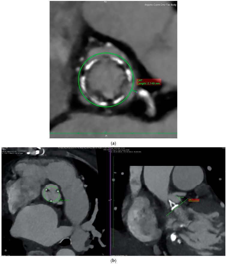
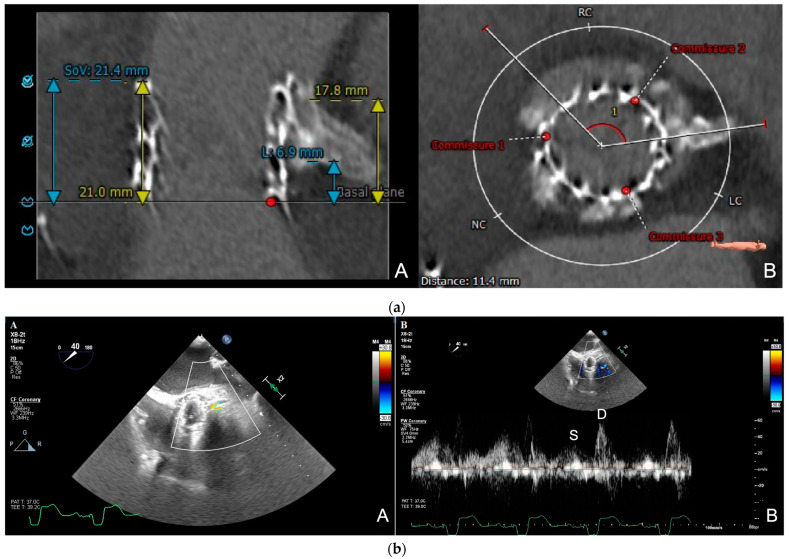
Valve-in-valve (ViV) transcatheter aortic valve replacement (TAVR) is emerging as an effective treatment for patients with symptomatically failing bioprosthetic valves and a high prohibitive surgical risk; a longer life expectancy has led to a higher demand for these valve reinterventions due to the increased possibilities of outliving the bioprosthetic valve's durability. Coronary obstruction is the most feared complication of valve-in-valve (ViV) TAVR; it is a rare but life-threatening complication and occurs most frequently at the left coronary artery ostium. Accurate pre-procedural planning, mainly based on cardiac computed tomography, is crucial to determining the feasibility of a ViV TAVR and to assessing the anticipated risk of a coronary obstruction and the eventual need for coronary protection measures. Intraprocedurally, the aortic root and a selective coronary angiography are useful for evaluating the anatomic relationship between the aortic valve and coronary ostia; transesophageal echocardiographic real-time monitoring of the coronary flow with a color Doppler and pulsed-wave Doppler is a valuable tool that allows for a determination of real-time coronary patency and the detection of asymptomatic coronary obstructions. Because of the risk of developing a delayed coronary obstruction, the close postprocedural monitoring of patients at a high risk of developing coronary obstructions is advisable. CT simulations of ViV TAVR, 3D printing models, and fusion imaging represent the future directions that may help provide a personalized lifetime strategy and tailored approach for each patient, potentially minimizing complications and improving outcomes.
瓣中瓣(ViV)经导管主动脉瓣置换术(TAVR)正逐渐成为治疗有症状的生物瓣膜功能衰竭且手术风险极高患者的有效方法;由于生物瓣膜耐久性的限制,患者预期寿命延长导致对这些瓣膜再次干预的需求增加。冠状动脉阻塞是瓣中瓣(ViV)TAVR最令人担忧的并发症;它是一种罕见但危及生命的并发症,最常发生在左冠状动脉开口处。准确的术前规划,主要基于心脏计算机断层扫描,对于确定ViV TAVR的可行性、评估冠状动脉阻塞的预期风险以及最终对冠状动脉保护措施的需求至关重要。在手术过程中,主动脉根部造影和选择性冠状动脉造影有助于评估主动脉瓣与冠状动脉开口之间的解剖关系;经食管超声心动图用彩色多普勒和脉冲波多普勒对冠状动脉血流进行实时监测是一种有价值的工具,可用于确定实时冠状动脉通畅情况并检测无症状冠状动脉阻塞。由于存在发生延迟性冠状动脉阻塞的风险,建议对有冠状动脉阻塞高风险的患者进行密切的术后监测。ViV TAVR的CT模拟、3D打印模型和融合成像代表了未来的发展方向,可能有助于为每位患者提供个性化的终身策略和量身定制的方法,潜在地减少并发症并改善治疗结果。