• 文献检索
  • 文档翻译
  • 深度研究
  • 学术资讯
  • Suppr Zotero 插件Zotero 插件
  • 邀请有礼
  • 套餐&价格
  • 历史记录
应用&插件
Suppr Zotero 插件Zotero 插件浏览器插件Mac 客户端Windows 客户端微信小程序
定价
高级版会员购买积分包购买API积分包
服务
文献检索文档翻译深度研究API 文档MCP 服务
关于我们
关于 Suppr公司介绍联系我们用户协议隐私条款
关注我们

Suppr 超能文献

核心技术专利:CN118964589B侵权必究
粤ICP备2023148730 号-1Suppr @ 2026

文献检索

告别复杂PubMed语法,用中文像聊天一样搜索,搜遍4000万医学文献。AI智能推荐,让科研检索更轻松。

立即免费搜索

文件翻译

保留排版,准确专业,支持PDF/Word/PPT等文件格式,支持 12+语言互译。

免费翻译文档

深度研究

AI帮你快速写综述,25分钟生成高质量综述,智能提取关键信息,辅助科研写作。

立即免费体验

足踝部软组织肿块的影像学评估:磁共振成像特征

Review of Soft Tissue Masses of the Foot and Ankle: Magnetic Resonance Imaging Features.

作者信息

Hegazi Tarek M, Aljamaan Yousef M, Alghamdi Shahad G, Alsaygh Jaffar S, Awary Khaled B, Aladel Fouad I, Elazomy Mohamed R, Almousa Sulaiman A

机构信息

Department of Radiology, Imam Abdulrahman Bin Faisal University, Dammam, Saudi Arabia.

Orthopedic Surgery, College of Medicine, King Fahd Hospital of the University, Imam Abdulrahman Bin Faisal University, Dammam, Saudi Arabia.

出版信息

Saudi J Med Med Sci. 2023 Apr-Jun;11(2):117-125. doi: 10.4103/sjmms.sjmms_66_22. Epub 2023 Apr 12.

DOI:10.4103/sjmms.sjmms_66_22
PMID:37252018
原文链接:https://pmc.ncbi.nlm.nih.gov/articles/PMC10211414/
Abstract

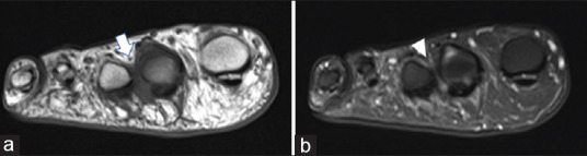
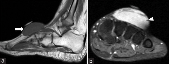
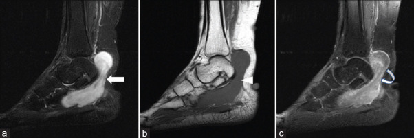
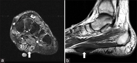
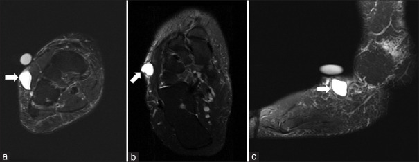
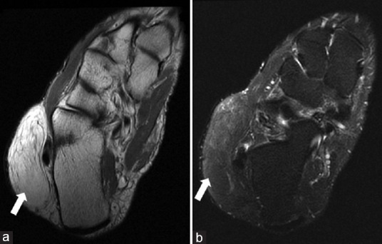
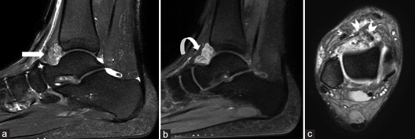
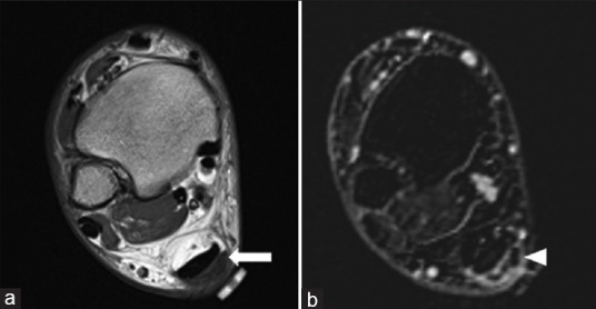
A large number of soft tissue masses affect the foot and ankle, with the majority being benign. Benign and malignant soft tissue lesions usually present as lumps, and it is important to differentiate between them to allow for optimal management. Imaging, in particular magnetic resonance imaging (MRI), can contribute to narrow the differential diagnosis of soft tissue masses of the foot and ankle by describing its exact location, internal signal characteristics, presence of enhancement, and its relation to adjacent structures. In this review, we review the literature to describe the most common soft tissue masses around the foot and ankle, focusing on the MRI features of the lesions.

摘要

大量软组织肿块会累及足踝部,其中大多数为良性。良性和恶性软组织病变通常表现为肿块,区分它们对于实现最佳治疗很重要。影像学检查,尤其是磁共振成像(MRI),通过描述软组织肿块的确切位置、内部信号特征、强化情况及其与相邻结构的关系,有助于缩小足踝部软组织肿块的鉴别诊断范围。在本综述中,我们回顾文献以描述足踝周围最常见的软组织肿块,重点关注这些病变的MRI特征。

https://cdn.ncbi.nlm.nih.gov/pmc/blobs/73c6/10211414/747f7e32b224/SJMMS-11-117-g010.jpg
https://cdn.ncbi.nlm.nih.gov/pmc/blobs/73c6/10211414/8307b57d4c91/SJMMS-11-117-g001.jpg
https://cdn.ncbi.nlm.nih.gov/pmc/blobs/73c6/10211414/2274eca71ff9/SJMMS-11-117-g002.jpg
https://cdn.ncbi.nlm.nih.gov/pmc/blobs/73c6/10211414/02c9757089a3/SJMMS-11-117-g003.jpg
https://cdn.ncbi.nlm.nih.gov/pmc/blobs/73c6/10211414/1647435abad9/SJMMS-11-117-g004.jpg
https://cdn.ncbi.nlm.nih.gov/pmc/blobs/73c6/10211414/fbf4ba8d78c3/SJMMS-11-117-g005.jpg
https://cdn.ncbi.nlm.nih.gov/pmc/blobs/73c6/10211414/fbf978e49d37/SJMMS-11-117-g006.jpg
https://cdn.ncbi.nlm.nih.gov/pmc/blobs/73c6/10211414/bb1b32dbdb69/SJMMS-11-117-g007.jpg
https://cdn.ncbi.nlm.nih.gov/pmc/blobs/73c6/10211414/76df4c68d0b2/SJMMS-11-117-g008.jpg
https://cdn.ncbi.nlm.nih.gov/pmc/blobs/73c6/10211414/72cc13d4e07e/SJMMS-11-117-g009.jpg
https://cdn.ncbi.nlm.nih.gov/pmc/blobs/73c6/10211414/747f7e32b224/SJMMS-11-117-g010.jpg

相似文献

1
Review of Soft Tissue Masses of the Foot and Ankle: Magnetic Resonance Imaging Features.足踝部软组织肿块的影像学评估:磁共振成像特征
Saudi J Med Med Sci. 2023 Apr-Jun;11(2):117-125. doi: 10.4103/sjmms.sjmms_66_22. Epub 2023 Apr 12.
2
MR Imaging of Common Soft Tissue Masses in the Foot and Ankle.足踝部常见软组织肿块的磁共振成像
Magn Reson Imaging Clin N Am. 2017 Feb;25(1):159-181. doi: 10.1016/j.mric.2016.08.013.
3
Benign and malignant tumors of the foot and ankle.足踝部的良性和恶性肿瘤
Skeletal Radiol. 2016 Mar;45(3):287-305. doi: 10.1007/s00256-015-2278-2. Epub 2015 Nov 4.
4
Soft tissue masses in the foot and ankle: characteristics on MR Imaging.足踝部软组织肿块:磁共振成像特征
Semin Musculoskelet Radiol. 2005 Sep;9(3):227-42. doi: 10.1055/s-2005-921942.
5
MRI imaging of soft tissue tumours of the foot and ankle.足踝部软组织肿瘤的磁共振成像
Insights Imaging. 2019 Jun 3;10(1):60. doi: 10.1186/s13244-019-0749-z.
6
Lumps and bumps around the foot and ankle: an assessment of frequency with ultrasound and MRI.足部和踝部的肿块和隆起:超声和 MRI 的评估频率。
Skeletal Radiol. 2013 Aug;42(8):1051-60. doi: 10.1007/s00256-013-1575-x. Epub 2013 Feb 6.
7
[Soft tissue tumors and pseudotumors of the foot and ankle].[足踝部软组织肿瘤及肿瘤样病变]
J Radiol. 2008 Jan;89(1 Pt 1):21-34. doi: 10.1016/s0221-0363(08)70366-4.
8
[Foot and ankle tumours : Part II: Malignant bone tumours and soft tissue tumours of the foot with differential diagnostic hints].[足踝部肿瘤:第二部分:足部恶性骨肿瘤及软组织肿瘤与鉴别诊断提示]
Radiologe. 2018 May;58(5):459-475. doi: 10.1007/s00117-018-0361-0.
9
MR imaging of benign soft-tissue masses of the foot and ankle.足踝部良性软组织肿块的磁共振成像
Radiographics. 1998 Nov-Dec;18(6):1481-98. doi: 10.1148/radiographics.18.6.9821196.
10
Magnetic Resonance Imaging Evaluation of Musculoskeletal Diseases of Ankle and Foot.踝关节和足部肌肉骨骼疾病的磁共振成像评估
Kathmandu Univ Med J (KUMJ). 2018;16(61):28-34.

本文引用的文献

1
An epidemiological review of 623 foot and ankle soft tissue tumours and pseudo-tumours.623 例足部和踝关节软组织肿瘤和假瘤的流行病学回顾。
Foot Ankle Surg. 2021 Jun;27(4):400-404. doi: 10.1016/j.fas.2020.05.004. Epub 2020 May 27.
2
MRI imaging of soft tissue tumours of the foot and ankle.足踝部软组织肿瘤的磁共振成像
Insights Imaging. 2019 Jun 3;10(1):60. doi: 10.1186/s13244-019-0749-z.
3
Malignant tumours of the foot and ankle.足部和踝关节的恶性肿瘤。
Foot Ankle Surg. 2020 Jun;26(4):363-370. doi: 10.1016/j.fas.2019.05.005. Epub 2019 May 11.
4
Current Concepts Review: Plantar Fibromatosis.当前概念综述:足底纤维瘤病
Foot Ankle Int. 2018 Jun;39(6):751-757. doi: 10.1177/1071100718768051. Epub 2018 Apr 5.
5
MR Imaging of Common Soft Tissue Masses in the Foot and Ankle.足踝部常见软组织肿块的磁共振成像
Magn Reson Imaging Clin N Am. 2017 Feb;25(1):159-181. doi: 10.1016/j.mric.2016.08.013.
6
Soft Tissue Tumors in Adults: ESSR-Approved Guidelines for Diagnostic Imaging.成人软组织肿瘤:ESSR批准的诊断成像指南
Semin Musculoskelet Radiol. 2015 Dec;19(5):475-82. doi: 10.1055/s-0035-1569251. Epub 2015 Dec 22.
7
Benign and malignant tumors of the foot and ankle.足踝部的良性和恶性肿瘤
Skeletal Radiol. 2016 Mar;45(3):287-305. doi: 10.1007/s00256-015-2278-2. Epub 2015 Nov 4.
8
Plantar fibromatosis--topical review.足底纤维瘤病——专题综述。
Foot Ankle Int. 2013 Dec;34(12):1742-6. doi: 10.1177/1071100713505535. Epub 2013 Sep 16.
9
Radiology-pathology conference: cutaneous angiosarcoma of the leg.放射科-病理科联合会诊:腿部皮肤血管肉瘤。
Clin Imaging. 2013 May-Jun;37(3):602-7. doi: 10.1016/j.clinimag.2012.08.005. Epub 2012 Sep 13.
10
Lumps and bumps around the foot and ankle: an assessment of frequency with ultrasound and MRI.足部和踝部的肿块和隆起:超声和 MRI 的评估频率。
Skeletal Radiol. 2013 Aug;42(8):1051-60. doi: 10.1007/s00256-013-1575-x. Epub 2013 Feb 6.