Girão Marília Maria Vasconcelos, Miyahara Lucas Kenzo, Dwan Viviane Sayuri Yamachira, Baptista Eduardo, Taneja Atul Kumar, Gotfryd Alberto, do Amaral E Castro Adham
Hospital Israelita Albert Einstein, São Paulo, Brazil.
Federal University of São Paulo, Rua Napoleão de Barros, n° 800, São Paulo, 04024-002, Brazil.
Insights Imaging. 2023 Jun 6;14(1):103. doi: 10.1186/s13244-023-01447-0.
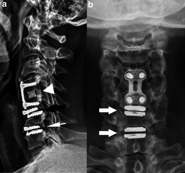
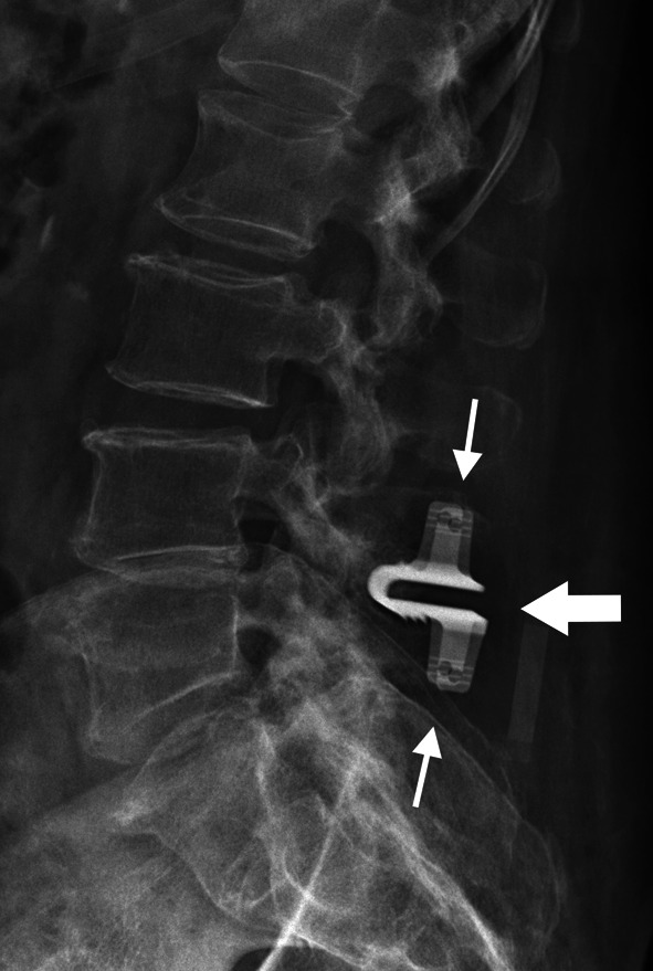
Spinal surgical procedures are becoming more common over the years, and imaging studies can be requested in the postoperative setting, such as a baseline study when implants are used, or when there is a new postoperative issue reported by the patient or even as routine surveillance. Therefore, it helps the surgeon in the appropriate management of cases. In this context, there is increasing importance of the radiologist in the adequate interpretation of postoperative images, as well as in the choice of the most appropriate modality for each case, especially among radiographs, computed tomography, magnetic resonance imaging and nuclear medicine. It is essential to be familiar with the main types of surgical techniques and imaging characteristics of each one, including the type and correct positioning of hardware involved, to differentiate normal and abnormal postoperative appearances. The purpose of this pictorial essay is to illustrate and discuss the more frequently used spine surgical interventions and their imaging characteristics, with an emphasis on classical decompression and fusion/stabilization procedures. KEY POINTS: Plain radiographs remain the main modality for baseline, dynamic evaluation, and follow-ups. CT is the method of choice for assessing bone fusion, hardware integrity and loosening. MRI should be used to evaluate bone marrow and soft tissue complications. Radiologists should be familiar with most performed spinal procedures in order to differentiate normal and abnormal. CRITICAL RELEVANCE STATEMENT: This article discusses the main surgical procedures involved in the spine, which can be didactically divided into decompression, stabilization-fusion, and miscellaneous, as well as the role of diagnostic imaging methods and their main findings in this context.
多年来,脊柱外科手术越来越普遍,术后可能需要进行影像学检查,例如在使用植入物时进行基线检查,或者患者报告有新的术后问题时,甚至作为常规监测。因此,这有助于外科医生对病例进行适当管理。在这种情况下,放射科医生对术后影像的充分解读以及为每个病例选择最合适的检查方式(尤其是在X线平片、计算机断层扫描、磁共振成像和核医学之间进行选择)变得越来越重要。必须熟悉主要的手术技术类型及其各自的影像学特征,包括所涉及硬件的类型和正确定位,以区分正常和异常的术后表现。这篇图文并茂的文章旨在阐述和讨论更常用的脊柱外科手术及其影像学特征,重点是经典的减压和融合/稳定手术。要点:X线平片仍然是基线、动态评估和随访的主要检查方式。CT是评估骨融合、硬件完整性和松动情况的首选方法。MRI应用于评估骨髓和软组织并发症。放射科医生应熟悉大多数脊柱手术,以便区分正常和异常情况。关键相关性声明:本文讨论了脊柱相关的主要手术操作,这些操作在教学上可分为减压、稳定融合和其他类型,以及诊断成像方法在此背景下的作用及其主要发现。